



- Medically Reviewed by: Mr Mfazo Hove, Consultant Ophthalmic Surgeon
- Author: Mr Mfazo Hove
- Published: November 19, 2024
- Last Updated: July 16, 2026
In short. Central retinal vein occlusion (CRVO) is a blockage of the main vein draining the retina, causing sudden, painless loss of vision in one eye. It is the more serious form of retinal vein occlusion, but modern anti-VEGF treatment offers a real chance of preserving, and often improving, vision. Prompt specialist assessment and ongoing monitoring matter.
The Blue Fin Vision® Rapid Medical Retina Pathway
- Prompt consultant-led assessment
- OCT imaging
- Diagnosis: wet AMD, dry AMD, RVO, CRVO or another retinal cause
- Intravitreal injection where appropriate
- Communication with your GP and optometrist on blood pressure, diabetes and vascular risk
- Ongoing monitoring, with escalation to Professor Mahmut Dogramaci, Consultant Vitreoretinal Surgeon, where needed
What Central Retinal Vein Occlusion Is, and Its Symptoms
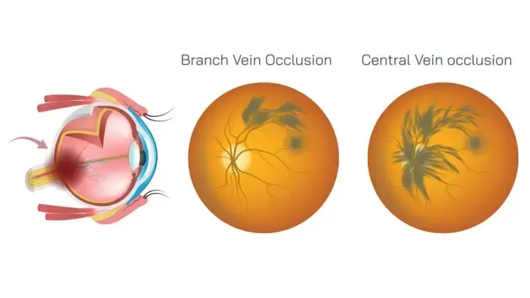
In CRVO, the central vein that drains the whole retina becomes blocked, so blood and fluid back up across the retina¹. This usually causes sudden, painless loss of vision in one eye. CRVO is the more severe form of retinal vein occlusion and needs specialist assessment².
Central Retinal Vein Occlusion Causes
CRVO is a thrombosis of the central retinal vein at the point where it leaves the eye through the optic nerve¹. It is closely linked to the health of the blood vessels, so the usual risk factors are high blood pressure, diabetes, raised cholesterol, glaucoma and increasing age³. In younger patients, clotting and inflammatory conditions are considered. CRVO is less common than branch vein occlusion but tends to be more serious⁴.
Two Types: Ischaemic and Non-Ischaemic
CRVO is divided into non-ischaemic (more common, with better retinal blood flow and usually better vision) and ischaemic (where widespread small-vessel closure leads to poorer vision and a higher risk of new-vessel complications)². The distinction drives both the outlook and the monitoring plan, and non-ischaemic CRVO can convert to the ischaemic type over time, so regular review matters⁵.
Complications We Monitor For
Two complications matter most. The first is macular oedema, swelling at the centre of vision, which is the main cause of blurring and is treatable¹. The second is the growth of abnormal new blood vessels (neovascularisation), including on the iris and drainage angle, which can raise the eye pressure and threaten sight if untreated². Careful, proactive monitoring is central to CRVO care, because catching these early allows prompt treatment.
Natural-history studies help set expectations. Without treatment, up to a third of non-ischaemic cases convert to the ischaemic type within three years, and among ischaemic eyes a significant proportion develop neovascular glaucoma within about 15 months⁵. Final vision is strongly influenced by the vision at presentation, which is why early assessment and treatment matter⁶.
Seek urgent review
Severe pain, nausea, a red eye or a sudden major rise in eye pressure after CRVO symptoms may suggest neovascular glaucoma and needs urgent ophthalmic assessment.
Why follow-up is not optional in CRVO
Even if the macular swelling improves, CRVO can lead to abnormal new blood vessels and a rise in eye pressure. Follow-up checks may include vision, OCT, eye pressure, pupil examination and, where needed, angiography or laser (panretinal photocoagulation).

Diagnosis of Central Retinal Vein Occlusion
Diagnosis uses a clinical examination and OCT, a quick, painless scan that measures macular swelling and guides treatment. Where further detail on the retinal circulation is needed, fluorescein angiography may be arranged. At Blue Fin Vision®, rapid-access OCT means change is detected and acted on quickly.
Central Retinal Vein Occlusion Treatment, With Real Hope
The outlook for CRVO has improved substantially. Anti-VEGF injections reduce macular oedema and improve vision in many patients⁷, and laser treatment is used where abnormal new vessels develop². Treatment is usually ongoing, with monitoring to protect vision over time. Complex vitreoretinal care and escalation are led by Professor Mahmut Dogramaci, Consultant Vitreoretinal Surgeon. Injections are delivered at The Harley Street Eye Centre, our dedicated London setting for private intravitreal injection care, with selected hospital sites available where clinically appropriate.
How Many Injections Might I Need?
CRVO usually needs sustained treatment, particularly in the first year. After initial monthly injections, the interval is extended as the swelling settles, using a treat-and-extend approach⁷. As a realistic guide, many people need in the region of 7 to 10 injections in the first year and fewer in the second, so often around 10 to 15 over two years, though some eyes need continued regular treatment. Where new vessels develop, laser (panretinal photocoagulation) is added to protect the eye⁶. Your consultant gives you a tailored estimate once your eye’s pattern is clear.
Support and Driving
If vision in one eye is affected, low vision support services can help you adapt, and your specialist can advise on this. Do not drive if your vision is suddenly reduced, distorted or unsafe. Many people with one affected eye can continue driving if the other eye meets DVLA standards, but the rules depend on whether one or both eyes are affected and whether the legal eyesight standard is met. Ask your specialist and check current DVLA guidance if unsure.
Frequently Asked Questions
Is CRVO worse than branch RVO?
It tends to be more serious, because the whole retina is affected rather than one part². Prompt, ongoing care gives the best outcome.
Can my vision improve?
Many patients improve with anti-VEGF treatment of the macular swelling⁷, though the degree of recovery varies with severity.
Why do I need regular follow-up?
To catch complications such as new vessel growth early², when they are most treatable. Monitoring is a core part of CRVO care.
Can I still drive with CRVO?
Often yes, if your other eye meets DVLA standards, but not while your vision is suddenly reduced or distorted. If both eyes are affected or you are unsure, check DVLA guidance and ask your specialist.
Will it affect my other eye?
The other eye is usually unaffected, but because CRVO is linked to blood-vessel health, managing blood pressure and related factors helps protect it³.
For sudden vision loss in one eye, or to discuss CRVO monitoring and treatment, book a prompt medical retina assessment.
Related Pages
References
- Wong TY, Scott IU. Retinal vein occlusion. N Engl J Med. 2010;363(22):2135-2144.
- Scott IU, Campochiaro PA, Newman NJ, Biousse V. Retinal vascular occlusions. Lancet. 2020;396(10266):1927-1940.
- Song P, Xu Y, Zha M, Zhang Y, Rudan I. Global epidemiology of retinal vein occlusion: a systematic review and meta-analysis of prevalence, incidence, and risk factors. J Glob Health. 2019;9(1):010427.
- Rogers SL, McIntosh RL, Cheung N, Lim L, Wang JJ, Mitchell P, Kowalski JW, Nguyen H, Wong TY. The prevalence of retinal vein occlusion: pooled data from population studies from the United States, Europe, Asia, and Australia. Ophthalmology. 2010;117(2):313-319.e1.
- McIntosh RL, Rogers SL, Lim L, Cheung N, Wang JJ, Mitchell P, Kowalski JW, Nguyen HP, Wong TY. Natural history of central retinal vein occlusion: an evidence-based systematic review. Ophthalmology. 2010;117(6):1113-1123.e15.
- Central Vein Occlusion Study Group. Natural history and clinical management of central retinal vein occlusion. Arch Ophthalmol. 1997;115(4):486-491.
- Brown DM, Campochiaro PA, Singh RP, Li Z, Gray S, Saroj N, Rundle AC, Rubio RG, Murahashi WY; CRUISE Investigators. Ranibizumab for macular edema following central retinal vein occlusion: six-month primary end point results of a phase III study. Ophthalmology. 2010;117(6):1124-1133.
ABOUT THE AUTHOR
Mr Mfazo Hove
Consultant Ophthalmic Surgeon
MBChB MD FRCOphth CertLRS
Mr Mfazo Hove is a Consultant Ophthalmic Surgeon with experience spanning more than 57,000 procedures. He completed 6.5 years of specialist training at Moorfields Eye Hospital and served for five years as a consultant at the Western Eye Hospital, Imperial College Healthcare NHS Trust. He is the founder of Blue Fin Vision®, a consultant-led private ophthalmology practice operating across London, Essex, and Hertfordshire. His clinical expertise encompasses advanced cataract surgery, refractive lens replacement, laser vision correction, and implantable Collamer lenses (ICL).
A ZEISS Key Opinion Leader, Mr Hove is a respected international speaker with five invited engagements across seven cities in 2026:
- ZEISS China tour (Changsha, Shanghai, and Hangzhou, April – ZEISS APAC User Meeting)
- RCOphth Annual Congress – May – Manchester
- ZEISS EMEA User Meeting (Istanbul)
- ZEISS Lausanne User Meeting (Lausanne)
- European Society of Cataract and Refractive Surgeons Annual Congress (ESCRS, London)


