




- Medically Reviewed by: Mr Mfazo Hove, Consultant Ophthalmic Surgeon
- Author: Mr Mfazo Hove
- Published: November 18, 2024
- Last Updated: November 5, 2026
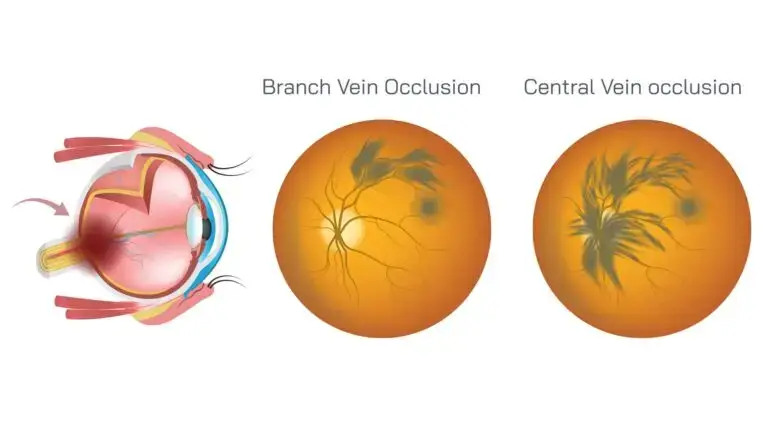
TL;DR: A branch retinal vein occlusion (BRVO) is a blockage in one of the smaller veins draining the retina, the light-sensitive layer at the back of the eye, which can blur or distort vision. The main threat to sight is swelling at the macula, the central part of the retina responsible for detailed vision, and this can be treated with injections. BRVO is also a reason to check your blood pressure and general circulation.
A blocked vein in the eye is often a message about the circulation everywhere else.
How Our Medical Retina Service Works
Blue Fin Vision® provides rapid access to assessment and treatment, followed by an organised next step.
Access
Contact the clinic, and we arrange a consultant consultation and optical coherence tomography (OCT) scan, typically the same day at the nearest suitable site. OCT produces detailed cross-sectional images of the retina.
Treatment
Where an injection is indicated, it is given at our Harley Street clinic the same day or the following day.
Continuity
Your next appointment is at four weeks, either with us or through an agreed NHS pathway.
We write to your NHS team, GP and optometrist so the handover is clear. If NHS treatment cannot be arranged in time, we can give the next injection.
Your Circumstances
Anti-vascular endothelial growth factor (anti-VEGF) medicines reduce leakage from retinal blood vessels. They are available on the NHS, and most people continue long-term treatment there; we often provide the first treatment and bridge to NHS care.
We also treat overseas patients and others who are not eligible for NHS-funded treatment. Medical retina consultations, OCT scans and injections are paid services.
For subsequent injections, we typically do not charge a separate consultation or OCT fee. If, at a follow-up visit, an injection is not needed, the consultation and OCT fees still apply.
Complication Readiness
We keep medication on hand to treat an acute rise in eye pressure after an injection. Every patient has the direct number of the consultant who performed their injection.
If pain, redness or worsening vision develops, even on a Sunday afternoon, call your consultant directly. We arrange urgent assessment, with vitreoretinal management, specialist care for the retina and the gel inside the eye, through Professor Mahmut Dogramaci where infection is suspected.
If you cannot reach your consultant promptly, do not wait: go to your nearest eye casualty or A&E department.
We plan for complications we have never had.
How Common It Is
Retinal vein occlusion is one of the most common retinal vascular conditions, which affect the blood vessels in the retina. Pooled population studies estimated that around 16 million adults worldwide are affected, most of them by branch rather than central occlusions. ¹
Why It Happens
A branch vein usually becomes blocked where a hardened artery crosses over it and compresses it. The main risk factors are: ²
- High blood pressure
- Diabetes
- Raised cholesterol
- Glaucoma or raised eye pressure
- Smoking and increasing age
Symptoms of Branch Retinal Vein Occlusion (BRVO)
- Sudden blurring or distortion in part of your vision.
- A shadow or patch, often in the upper or lower half of your sight.
- Sometimes no symptoms at all, if the blockage is away from the macula.
If you experience sudden loss or deterioration of vision, seek urgent assessment through an eye clinic or eye casualty service. Do not wait for a routine appointment. ⁸
How It Can Affect Sight
Macular Oedema
Macular oedema means swelling caused by fluid leaking into the macula. It is the most common cause of reduced vision in BRVO.
Anti-VEGF injections are now the main treatment. In the BRAVO trial, eyes treated with injections gained substantially more vision at six months than untreated eyes. ³
Before injections, grid laser, which applies small laser spots to selected areas of the retina, was shown to improve vision in some eyes with macular oedema. ⁴
New Vessels
Where large areas of retina lose their blood supply, fragile new vessels can grow and bleed into the vitreous, the clear gel inside the eye. Laser to the affected area reduces this risk. ⁵

What Happens Next
You will have OCT imaging to look for swelling, and your eye is monitored over the following months.
Your GP should check your blood pressure, blood sugar and cholesterol, because treating these protects both eyes and your general health.
Our articles explain the related conditions:
What Happens at Your Eye Clinic Appointment
Your eye clinic appointment includes checks of your vision, eye pressure and retinal health.
- Your vision and eye pressure are measured; raised eye pressure is a risk factor for vein occlusion.
- Your blood pressure is checked.
- Dilating drops widen your pupils to allow a full examination of the retina, where the blocked vein and haemorrhages, areas of bleeding, are usually visible.
- An OCT scan shows whether fluid has reached the macula.
- Further imaging may show how much of the retina has lost its blood supply. OCT angiography maps blood flow without an injected dye. Fluorescein angiography uses a dye injected into a vein in your arm and photographs to assess retinal circulation.
Blood tests through your GP, including blood sugar and cholesterol, look for underlying risk factors. Younger patients may need further tests. ²
Anti-VEGF Injections for BRVO
If fluid has reached the macula and reduced your vision, injections are usually given a month apart until the macula is dry, meaning the swelling has resolved, then spaced out as scans allow.
These are intravitreal injections, meaning the medicine is delivered into the vitreous gel inside the eye. Each injection takes seconds under numbing drops.
Mild grittiness, a red patch on the white of the eye or a few floaters, small shapes drifting across your vision, are common afterwards. Serious complications such as infection are rare, and you will be told which symptoms need urgent attention.
In the BRAVO trial, most of the improvement in vision came in the early months of treatment. ³
Other Treatments
- Steroid implants placed inside the eye can reduce swelling that persists despite injections.
- Laser can treat persistent macular swelling in selected eyes, and treats areas of retina without a blood supply if new vessels develop. ⁴ ⁵
- Vitrectomy surgery, an operation to remove the vitreous gel, is occasionally needed if bleeding into the vitreous does not clear.
What Is the Outlook?
Many people with BRVO keep good vision, particularly when the macula is not involved or swelling is treated promptly.
Some mild occlusions improve without treatment as the circulation re-routes. How much vision returns depends on how badly the macula was affected and for how long.
Protecting Your Other Eye and Your Health
The most important step is controlling the risk factors that caused the blockage: blood pressure, blood sugar, cholesterol and smoking. This reduces the chance of a vein occlusion in the other eye and protects your heart and brain.
Frequently Asked Questions
Is a vein occlusion a stroke in the eye?
Not exactly. A stroke usually involves an artery, while this is a vein. However, they share risk factors, so a vein occlusion is a good reason to have your general health checked.
Can it happen in the other eye?
It can, although most people only ever have one. Controlling blood pressure and other risk factors reduces the risk.
Will my vision come back?
Often, at least partly. Vision improves as swelling settles, either with treatment or on its own in milder cases.
How many injections will I need?
It varies. Some people need only a few; others need injections over a year or more. OCT scans guide the timing.
Do I need to stop my medicines?
Usually not. Keep taking your prescribed medicines, including blood thinners, unless your doctor advises otherwise.
Why We Plan for Infection
Serious infection inside the eye, called endophthalmitis, is rare after an injection.
In a regional study in England, the risk was around 1 in 4,000 injections, similar per procedure to cataract surgery. Because far more injections were performed, they accounted for more cases; those figures describe that population, not our own patients. ⁶
Intracameral antibiotics, antibiotics delivered into the front chamber of the eye, are standard infection prevention in cataract surgery. ⁷ Anti-VEGF injections do not provide that antibiotic protection, so meticulous prevention and a clear emergency response pathway remain essential.
Mr Hove led the Eye Casualty service at the Western Eye Hospital for five years.
We have never had a case of endophthalmitis after an injection; we plan for it anyway, so that if symptoms develop, assessment and treatment are not delayed.
At Blue Fin Vision®
Macular oedema from BRVO is treated under our Rapid Medical Retina Pathway, described above.
We write to your GP about vascular risk factors, conditions affecting the health of your blood vessels. If bleeding into the vitreous needs surgery, care is escalated to Professor Mahmut Dogramaci, Consultant Vitreoretinal Surgeon, within the same team.
Read more about our care and treatment:
- Our Medical Retina service.
- Our Private Intravitreal Injections.
- Our Patient Information explains more.
Book a consultation with Blue Fin Vision® to discuss your BRVO diagnosis, treatment and ongoing monitoring, with consultant-led care at clinics across London, Hertfordshire and Essex.
References
- Rogers S, McIntosh RL, Cheung N, Lim L, Wang JJ, Mitchell P, et al.; International Eye Disease Consortium. The prevalence of retinal vein occlusion: pooled data from population studies from the United States, Europe, Asia, and Australia. Ophthalmology. 2010;117(2):313-9.e1.
- Wong TY, Scott IU. Clinical practice. Retinal-vein occlusion. N Engl J Med. 2010;363(22):2135-44.
- Campochiaro PA, Heier JS, Feiner L, Gray S, Saroj N, Rundle AC, et al.; BRAVO Investigators. Ranibizumab for macular edema following branch retinal vein occlusion: six-month primary end point results of a phase III study. Ophthalmology. 2010;117(6):1102-12.e1.
- Branch Vein Occlusion Study Group. Argon laser photocoagulation for macular edema in branch vein occlusion. Am J Ophthalmol. 1984;98(3):271-82.
- Branch Vein Occlusion Study Group. Scatter photocoagulation for prevention of neovascularization and vitreous hemorrhage in branch vein occlusion. Arch Ophthalmol. 1986;104(1):271-82.
- Ong AY, Rigaudy A, Toufeeq S, Robins J, Shalchi Z, Bindra MS, et al. Intravitreal injections as a leading cause of acute postoperative endophthalmitis: a regional survey in England. Eye (Lond). 2023;37(1):163-9. doi:10.1038/s41433-021-01886-3.
- ESCRS Endophthalmitis Study Group. Prophylaxis of postoperative endophthalmitis following cataract surgery: results of the ESCRS multicenter study and identification of risk factors. J Cataract Refract Surg. 2007;33(6):978-88.
- Cornwall and Isles of Scilly Referral Management Service. Retina and vitreous [Internet]. NHS; [cited 2026 Nov 4].
ABOUT THE AUTHOR
Mr Mfazo Hove
MBChB MD FRCOphth CertLRS
Consultant Ophthalmic Surgeon | Founder and Medical Director, Blue Fin Vision®
Mr Mfazo Hove is a Consultant Ophthalmic Surgeon with more than two decades of experience across NHS and private practice and more than 57,000 ophthalmic procedures performed. He trained for six and a half years at Moorfields Eye Hospital and served for five years as an NHS consultant at the Western Eye Hospital, Imperial College Healthcare NHS Trust.
He specialises in cataract surgery, refractive lens replacement, laser vision correction and implantable Collamer lenses (ICL). He is the founder and Medical Director of Blue Fin Vision®, a private ophthalmology group providing consultant-delivered care across London, Hertfordshire and Essex. His cataract surgery outcomes are publicly reported through the National Ophthalmology Database.
A ZEISS Key Opinion Leader and international speaker, Mr Hove is a contributing author to The Ophthalmologist and Ophthalmology Times. His writing examines how clinical governance and the organisation of ophthalmic services shape patients’ experiences and outcomes, with a focus on transparent outcome reporting, surgical training and long-term accountability for patient care.


