




- Medically Reviewed by: Mr Mfazo Hove, Consultant Ophthalmic Surgeon
- Author: Mr Mfazo Hove
- Published: October 13, 2026
- Last Updated: October 10, 2026
In Brief
- Modern implantable collamer lenses (ICLs) with a central port, a tiny opening in the centre of the implant, have produced strong, stable vision over three to ten years in carefully selected patients.¹ ² ³
- Your natural lens stays in place, so your eyes keep ageing around the implant. Reading glasses in your forties and, eventually, cataract are part of that.⁴ ⁵
- Long-term follow-up checks the implant and the health of the eye. It is not simply another glasses test.
- At Blue Fin Vision®, annual vault measurements, which check the gap between the implant and your natural lens, and endothelial cell counts, which assess the cells lining the inside of the cornea, the clear front surface of the eye, are included at no additional cost for as long as your ICL remains in the eye, under our published ICL Lifetime Monitoring Protocol™.
- New symptoms should be assessed promptly, not saved for your next routine visit.⁶
“Lifetime care” describes our whole approach. The free monitoring commitment is precisely defined; it does not make every future eye treatment free.
The Implant Stays, So The Care Must Too
Most people considering ICL surgery start with two questions: how well will I see, and how quickly will I recover? Both matter. A third question matters just as much and is asked far less often: who will look after my eyes in five, ten or twenty years?
An ICL is a thin, flexible lens placed behind the iris (the coloured part of the eye) and in front of your natural lens, to correct short-sightedness, long-sightedness or astigmatism, blurred vision caused by uneven curvature of the cornea or natural lens. Clinicians call it a phakic lens, because your own lens (the “phakos”) stays in place. That is the key difference from cataract surgery and lens replacement, where the natural lens is removed and replaced.⁴ ⁷

An ICL sits behind the iris and in front of your natural lens, which stays in place.
The operation takes minutes. The implant stays for decades, and so does the clinical relationship that begins at your first consultation. Choosing an ICL therefore also means choosing a follow-up pathway. For many people with higher prescriptions or thinner corneas, the ICL is the better operation: an honest alternative to laser, not a consolation prize. This article explains what is measured after surgery, why those measurements are repeated even when vision feels perfect, and which decisions they help us make.
What Modern Long-Term Studies Show
The modern evidence is reassuring, and it is also the reason careful studies keep measuring. Three studies of central-port lenses illustrate both points.
Study | What It Followed | What It Reported |
|---|---|---|
United States Food and Drug Administration (FDA) prospective multicentre study, 2025¹ | 629 eyes of 327 people with EVO/EVO+ lenses, over three years | 90.7% of eyes examined at three years were within ±0.50 dioptres (D), a measure of prescription strength, of the target prescription. Mean endothelial cell density was 6.7% lower than before surgery. |
Japanese central-port study, 2021² | 177 eyes, with results extending to eight years | Vision remained stable. The three eyes that developed cataract all had small anterior subcapsular changes, early clouding just beneath the front of the natural lens, before surgery. |
Korean ten-year V4c study, 2025³ | 103 eyes followed for ten years | Vision remained stable, while both vault and endothelial cell density declined gradually over the decade. |
These figures are study results, not Blue Fin Vision® outcomes. The FDA study was sponsored by the lens manufacturer, and several of its authors declared financial relationships with it.¹
Three points help in reading them. A three-year result and a ten-year result answer different questions. The people studied, their starting prescriptions and how many returned for late visits all differ: in the eight-year study, roughly a third of eyes were examined at eight years.² And no study can show a lifetime risk of zero or predict what will happen in one person’s eye.
Lens design also matters. The central port, a tiny opening in the middle of the ICL, allows fluid to flow through the lens. In a five-year paired comparison, central-port and conventional lenses performed similarly, and only the conventional group recorded an anterior subcapsular cataract, clouding just beneath the front of the natural lens.⁸ Older studies of lenses without a port reported much higher rates of lens opacity, or clouding, over ten to twelve years.⁹ ¹⁰ Those figures describe earlier designs and are not a risk estimate for today’s EVO lens.

The EVO ICL. The tiny opening at the centre of the optic is the central port.
Monitoring is justified by the information it gives over time, not by any suggestion that modern ICL surgery often fails.
Lifetime Care Starts With The Preoperative Baseline
The measurements taken before surgery are the reference point for every later visit.
Your preoperative assessment records your prescription and best corrected vision, meaning your clearest vision with glasses or contact lenses, the depth and dimensions of the front chamber of the eye, endothelial cell density, eye pressure, the clarity of your natural lens, and the retina (the light-sensitive layer at the back of the eye) where your history or prescription calls for it.⁴ ⁷ A credible assessment must be capable of ending with a recommendation not to proceed, when another option or no surgery would serve you better.
Sizing the ICL is a clinical decision informed by measurement. Different imaging methods, such as anterior segment optical coherence tomography (AS-OCT), a scan that produces detailed images of the front of the eye, and high-frequency ultrasound, show different parts of the anatomy, and published sizing methods support careful assessment.¹¹ No single instrument or calculation makes sizing infallible, which is one reason the result is checked after surgery. At Blue Fin Vision®, sizing uses anterior segment OCT (ANTERION and REVO). We combine the manufacturer’s STAAR sizing calculation with an artificial intelligence (AI)-based formula, and the operating consultant reviews both before choosing the lens size. Using two methods gives a cross-check; it does not replace measuring the vault once the lens is in place.

The main measurements considered in ICL sizing. Schematic, not to scale.
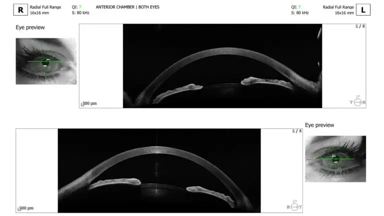
Anterior segment OCT of both eyes before surgery, used to measure the front of the eye for sizing.
Your record should also hold:
- The lens model, size and power, and the date of implantation
- The intended axis, or alignment, if you have a toric (astigmatism-correcting) ICL
- Your preoperative measurements, kept available if you move or later see another clinician
Early Aftercare and Long-Term Follow-Up Do Different Jobs
Early visits check recovery: inflammation, eye pressure, how the lens has settled and how well you see. Later visits look for change in measurements that were stable, and for new conditions in the eye. A rise in pressure soon after surgery can have several causes, including retained gel from the operation or a response to steroid drops, and it is managed differently from a pressure problem that appears years later.¹²
Your schedule at Blue Fin Vision® is the same for every ICL patient:
When | What Happens | Cost |
|---|---|---|
From the moment you leave theatre | You have the direct phone number of your operating consultant | Included |
Day after surgery | Your operating consultant speaks with you; you are examined that day if there is a clinical reason | Included |
Week 1 | First examination | Included in the surgery fee |
Months 1 and 3 | Consultant reviews; first endothelial cell count at month 3 | Included in the surgery fee |
Month 12 | Consultant review with vault imaging, endothelial cell count, eye pressure, slit-lamp examination (a microscope examination of the eye) and refraction (measurement of your prescription), and a written report | Included in the surgery fee |
Every year after that | The same tests and written report, for as long as the ICL remains in your eye: vault at any Blue Fin Vision® site, endothelial count at Harley Street | Included in the ICL Lifetime Monitoring Protocol™ |
Sooner, when needed | A new symptom or a change in your measurements; your consultant sets the interval | See what is included, below |
Endothelial cell counts are annual for as long as the ICL remains in your eye. After the initial postoperative period, they do not move to every two years at any point.
If anything changes between visits, you do not wait for the next appointment: a symptom on day two is assessed on day two.
Vault: Why The Space Around The Implant Matters
Vault is the gap between the back surface of the ICL and the front surface of your natural lens. You cannot feel it, so it is measured with imaging, not judged by how well you see.

Schematic of the front of the eye showing the vault, not to scale.
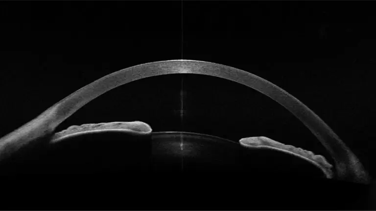
Before surgery: the natural lens sits directly behind the iris.


After surgery: the ICL shows as a thin bright line in front of the natural lens. The vault is measured at 694 micrometres (µm) and 662 µm in these two scans.
Both ends of the range matter. A low vault brings the implant closer to the natural lens, which in older lens designs was linked with lens opacity.⁹ With the current central-port lens, a ten-year study of eyes with low and normal vault found no lens opacity caused by the ICL, even in eyes below 250 µm.¹³ A high vault can push the iris forward and crowd the drainage angle, where fluid leaves the eye. That is why vault is always read alongside the angle, the eye pressure and the clarity of the natural lens.³ ¹⁴
Vault is not fixed for life. The natural lens slowly thickens with age, and the ten-year V4c study recorded a gradual decline in vault over time.³ One reading tells us where the lens sits today; a series shows how the gap changes over time.
At Blue Fin Vision®, a vault outside 250–750 µm on two successive measurements, or a significant drift from your own baseline, prompts a consultant review. This is our clinical reference range, not a regulatory one, and it is not an automatic trigger for surgery. What it means depends on your anatomy, your lens model, the other findings and the direction of change. The usual first steps are repeat imaging and closer observation. Repositioning, exchanging or removing the lens are discussed only when the overall picture supports it.
Endothelial Cells: Why Good Vision Is Not The Only Measure
The endothelium is the single layer of cells lining the inside of the cornea. It pumps fluid out of the cornea to keep it clear, and its cells do not regenerate. Gradual loss causes no symptoms until it is advanced.
The measurement is specular microscopy, which photographs the cells and reports endothelial cell density in cells per square millimetre (cells/mm²). Clinicians often shorten this to ECC, for endothelial cell count. It is measured at three months, at 12 months and then once a year, as shown in the schedule above.
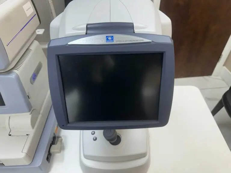
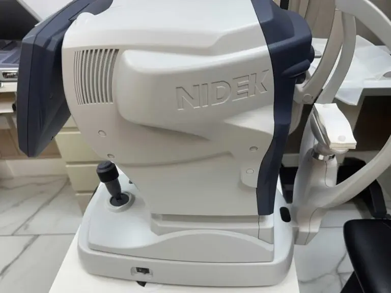
The specular microscope we use for endothelial cell counts.
A single number tells us less than a reliable series. A reading of 2,400 cells/mm² in an eye that measured 2,900 three years earlier means something different from the same reading in an eye that has held steady since surgery. That is why each monitoring visit ends with a written report against your own baseline. Each reading is interpreted against your age, your reserve before surgery, the quality of the image and the rest of the examination. An unexpected result is usually repeated before any decision is made.
Published studies show a range: modest average loss in several central-port cohorts, with individual eyes varying around that average.¹ ² ³ ¹⁵ In one paired-eye study, the eye with the higher vault tended to lose more cells than its fellow eye, an association rather than proof that vault was the cause.¹⁶ Monitoring does not stop cell loss. It shows when a pattern deserves attention.
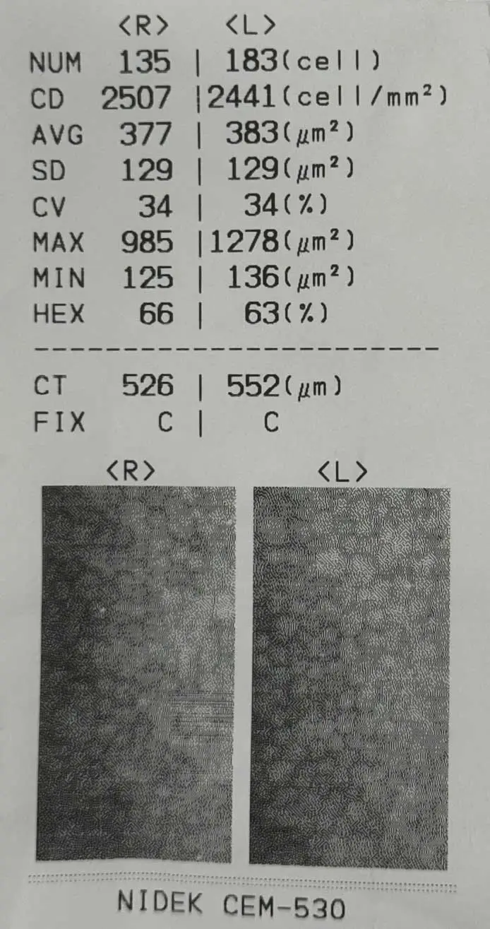
A specular microscopy report. CD is cell density: 2,507 cells/mm² in the right eye and 2,441 cells/mm² in the left. CT is central corneal thickness, in micrometres.
At Blue Fin Vision®, a density trending below 2,000 cells/mm², or a rate of loss steeper than expected for your age, prompts a consultant review. If loss looks clinically important, the pathway is to confirm the finding, look for a cause, shorten the interval between visits and discuss whether the implant should stay. Published series describe eyes in which the ICL was removed because of endothelial cell loss, which is why a trend is acted on rather than watched indefinitely.¹⁷
If cell loss were allowed to progress far enough, the cornea could swell and cloud, a condition called corneal decompensation. This is not an expected outcome of modern ICL surgery, and finding a trend early is the point of measuring. Can my cornea decompensate after having ICL surgery? covers it in more detail.
Eye Pressure and Glaucoma are Related but Different
Raised eye pressure is a measurement. Glaucoma is damage to the optic nerve, which carries visual information from the eye to the brain. A high reading on its own is not a diagnosis of glaucoma, and glaucoma can occur at normal pressures.¹² ¹⁸
That is why different tests answer different questions. A pressure check measures the pressure. Gonioscopy, a contact lens examination of the drainage angle, shows whether the angle is open or crowded. Optic nerve imaging (OCT) and visual field testing, which checks your central and peripheral vision, show whether the nerve is being affected. Those extra tests are carried out when the findings call for them, not at every annual visit.
Early pressure rises after surgery usually have different causes from problems that appear years later.¹² The central port improves fluid flow and has largely removed the need for the laser iridotomy that older ICLs required, a procedure that creates a small opening in the iris to help fluid circulate.⁸ In the three-year FDA study, no eye had raised pressure from pupillary block, a blockage of fluid flow through the pupil, or angle narrowing.¹ The port does not remove every cause of raised pressure, or the ordinary age-related risk of glaucoma, so pressure is checked at every monitoring visit. Can I get glaucoma after having ICL surgery? explains this further.
Cataract Is a Pathway, Not a Surprise
Keeping your natural lens means keeping the possibility of age-related cataract, as everyone does. A cataract after ICL surgery is not automatically caused by the ICL, and a small opacity in the lens is not the same as a cataract that affects your vision enough to justify surgery.⁵ ⁹ ¹⁰
When surgery is needed, the plan usually combines three steps: removing the ICL, removing the cloudy natural lens, and implanting a new intraocular lens (IOL), an artificial replacement lens inside the eye. These are often done in one operation, but the plan is individual. A five-centre Japanese series of 83 eyes found this combined approach practical, with good early visual results.¹⁹ It looked at selected cases over three months, included older and newer ICL designs, and cannot tell us how often cataract occurs.
The choice of replacement lens depends on your retina, cornea, measurements and priorities. Measurements for cataract surgery need careful checking because the existing ICL can affect how some devices identify the eye’s structures, so it is worth asking any clinic whether it has done this surgery before.
At Blue Fin Vision®, cataract care follows on within the same clinical team. The published Post-ICL Cataract Surgery Pathway covers ICL removal, cataract surgery and a standard monofocal (single-focus) or toric IOL, priced in advance per eye, as set out below. Premium lenses are an optional upgrade. What happens if I get cataracts after having ICL surgery? goes into more detail.
Time Since ICL Implantation | Cost Of Cataract Surgery, Per Eye |
|---|---|
Up to 2 years | No charge |
2 to 10 years | 50% of the Blue Fin Vision® standard fixed cataract surgery price |
After 10 years | The Blue Fin Vision® standard fixed cataract surgery price |
Correcting The Prescription Does Not Remove Retinal Risk
An ICL corrects how the eye focuses. It does not change the shape of a highly short-sighted eye, and a longer eye carries a higher background risk of retinal tears and detachment, when the retina pulls away from the back of the eye.⁶
Two long-term comparative studies found no significant difference in retinal detachment rates between eyes with an ICL and similar short-sighted eyes without surgery.²⁰ ²¹ That is reassuring context. It does not show that ICL surgery protects the retina, or that any individual is free of risk.
If you do develop a retinal problem, prompt access matters more than anything else. Blue Fin Vision® offers vitreoretinal assessment and surgery, care for the retina and the vitreous gel inside the eye, with seven-day emergency retinal capability led by Professor Mahmut Dogramaci, Consultant Vitreoretinal Surgeon. Treatment of retinal disease unrelated to the ICL is charged according to the treatment needed. Care for a complication of your ICL surgery follows the terms of your surgical agreement. See vitreoretinal surgery.
Why Vision Can Change Without the ICL “Wearing Out”
An ICL does not have an expiry date, and it is not guaranteed never to need removal. When vision changes, the cause is usually the eye around the lens, not the lens itself.
The common reasons are natural ageing and presbyopia, the age-related loss of near focusing ability, which means most people with an ICL need reading support from their mid-forties; a change in prescription; early cataract; or another eye condition.⁴ ⁵ An ICL corrects the prescription you have at the time of surgery. It does not stop short-sightedness from progressing.
With a toric ICL, residual or new astigmatism is not the same as lens rotation. Rotation is confirmed by examination before anything is done about it. Does ICL surgery correct astigmatism? explains the basics. Either way, a changed prescription calls for an assessment, not an assumption that another operation is needed.
Enhancement: When A Residual Prescription Is Worth Treating
Sometimes a small prescription remains after surgery. Whether to treat it is a clinical judgement, not an automatic step.
Blue Fin Vision® does not enhance below 1.00 dioptre of sphere (the short- or long-sighted part of your prescription) or cylinder (the astigmatism component). Reaching 1.00 D starts the conversation; it does not decide it. Your surgeon then asks three questions:
- Is the residual error large enough to matter?
- Is it actually causing your visual complaint?
- Does the likely benefit outweigh the risks of another procedure?
The answers draw on your vision, a stable refraction, corneal imaging and thickness, the ocular surface (the front surface of the eye and its tear film), the position of the ICL and the original target. Laser on the cornea and a change of implant are not interchangeable, and the right choice depends on why the error is there.
On the ICL pathway, eligible enhancement carries no additional fee: no surgeon, hospital, diagnostic or drops fee. It is normally completed within 12 months of surgery, and eligibility can extend to 24 months where a genuine need was identified within the pathway. Enhancements are carried out within Blue Fin Vision®, not outsourced. Enhancement cannot promise a zero prescription, freedom from haloes or that your eyes will not change again. Any potential enhancement or further treatment policy will be explained clearly during consultation, in writing, before you consent.
The full terms are in our Vision Enhancement Patient Information leaflet.
What Should a Long-Term ICL Review Assess?
A long-term review connects each test with a question about your eye. The core tests follow the schedule above. Additional investigations, such as angle, optic nerve or retinal imaging, depend on what the findings show, and your consultant will tell you before any test outside the ICL Lifetime Monitoring Protocol™ is arranged.
Assessment | Purpose |
|---|---|
Symptoms and visual function | Ask about changes, glare, near vision and daily activities |
Vault and lens position | Check where the implant sits and compare with earlier images |
Endothelial cell density | Review cell reserve and whether any trend is reliable |
Pressure and drainage angle | Measure pressure and decide whether angle or glaucoma tests are needed |
Natural lens and cornea | Look for early cataract and corneal change |
Retina and optic nerve | Examine according to symptoms, prescription, history and risk |
You should leave each review with three things: an explanation of your results, the date of your next review, and a clear way to reach us if something changes before then.
What To Ask Any Clinic Before Surgery
Because an ICL is intended to remain in the eye long term, these questions belong in your first consultation. Our ICL Surgery Checklist has the full list; these are the ones about long-term care:
- What is my endothelial cell count before surgery?
- How often will vault and endothelial cell density be measured, and is that included in the price?
- What happens if a measurement changes?
- Who performs an enhancement if I need one, and on what terms?
- Who holds my records if I move or live abroad?
It is reasonable to ask for the answers in writing.
What Blue Fin Vision® Means By Lifetime ICL Care
Every patient who has ICL surgery at Blue Fin Vision® is enrolled in the ICL Lifetime Monitoring Protocol™ at the point of surgery. There is no subscription, no separate fee and no expiry. Annual vault measurements and endothelial cell counts are included at no additional cost for as long as the ICL remains in your eye. The manufacturer’s labelling also advises continued endothelial monitoring for as long as the lens remains implanted.²²
What Is Included
- All scheduled monitoring visits from month 12 onwards
- Specular microscopy, AS-OCT and Pentacam imaging, which maps the shape and thickness of the cornea
- Eye pressure, slit-lamp examination and refraction at every monitoring visit
- A written report after each visit, comparing your results with your own baseline and previous years
- A consultant review whenever a measurement crosses a defined threshold or shows a clinically significant change
What Is Not Included
Appointments you request for reasons unrelated to your monitoring results, such as a general eye check or a second opinion, which are charged at standard consultation fees; treatment of unrelated conditions such as glaucoma, macular disease (conditions affecting the central part of the retina) or diabetic eye disease; refractive error unrelated to the original ICL; contact lens fitting; and travel. New symptoms are assessed according to clinical urgency. Any applicable charges are explained in advance. The full terms are on the ICL Lifetime Monitoring Protocol™ page.
How Serial Results Are Used
Your consultant reviews every measurement on a single timeline. If treatment is needed, ICL exchange, ICL removal and cataract surgery are all carried out within Blue Fin Vision®. Their costs are set out in the cataract and enhancement sections above.
Where Monitoring Takes Place
Vault measurements are performed at Blue Fin Vision® sites, including Harley Street, Weymouth Street, Chase Lodge Hospital, Phoenix Hospital Chelmsford and One Hatfield Hospital. Endothelial cell counts are performed at Harley Street, so if you usually attend a regional clinic, your annual endothelial count still takes place in Harley Street. All monitoring takes place at Blue Fin Vision® clinics in the UK, so if you live abroad or far from our sites, plan to return for your annual visits, or have the same tests done where you are, at your own cost.
If your ICL was implanted elsewhere, you are welcome to have it assessed through a separate paid assessment pathway. Enrolment in the ICL Lifetime Monitoring Protocol™ at no additional cost applies to ICLs implanted at Blue Fin Vision®.
Symptoms That Should Not Wait For Your Next Review
Seek urgent assessment the same day if you notice:
- Sudden loss of vision or marked new blurring
- New flashes of light or a shower of new floaters
- A curtain or shadow across part of your vision
- Significant eye pain or redness
- Severe eye pain with headache, nausea or haloes around lights⁶ ¹⁸
Blue Fin Vision® patients: call your operating consultant on the direct number you were given at surgery. During clinic hours you can also call 07704 225640 (Monday to Friday, 10am to 6pm).
If you cannot reach us, go to your nearest eye casualty or A&E, or call NHS 111. Do not wait for a reply to an online booking request.
Frequently Asked Questions
Do I still need checks if I see perfectly?
Yes. Clear vision tells us the optics are working. It does not show your vault, your endothelial cell density or your eye pressure, and each can change without symptoms.
Is an annual review enough for everyone?
Annual review is our usual framework for stable eyes; your consultant individualises the interval. A borderline measurement or a new symptom brings the next visit forward.
Does the EVO central port remove the need for follow-up?
No. The port solved specific problems seen with older lenses. It does not prevent age-related change in the natural lens, gradual cell loss or unrelated eye disease.
Will my ICL need replacing after a fixed number of years?
No. There is no set expiry. A lens is exchanged or removed only when the measurements or your vision give a clinical reason.
What if I develop cataracts?
Your consultant assesses whether the cataract is affecting your vision. If surgery is needed, the ICL is removed and the cataract treated, usually in one operation, under the published Post-ICL Cataract Surgery Pathway.
Does lifetime monitoring mean every future treatment is free?
No. The ICL Lifetime Monitoring Protocol™ covers defined monitoring and threshold-triggered consultant review. Cataract surgery after ICL follows its own published pricing. Eligible enhancement is covered under its own policy; other treatments may carry separate charges. The terms are written down before your surgery.
Service Documentation
These documents describe our service commitments and are not independent outcome evidence:
- Blue Fin Vision®. ICL Lifetime Monitoring Protocol™. Service information. Accessed 7 October 2026.
- Blue Fin Vision®. Implantable Collamer Lens Surgery at Blue Fin Vision®. Service information. Accessed 7 October 2026.
- Blue Fin Vision®. ICL Surgery Patient Information. Version 1.0, August 2026.
- Blue Fin Vision®. ICL Surgery Checklist.
- Blue Fin Vision®. Vision Enhancement Patient Information. Version 1.0, August 2026.
References
- Parkhurst G, Brinton JP, Faulkner A, Moshirfar M, Kugler LJ, Stahl JE, Zavodni Z, Thompson VM, Price FW, Wiley WF, Aronsky M, Whitman JD, Solomon JD, Perkins SA, Packer M, Egamino J. Three year results from the United States FDA prospective multicenter clinical study of the EVO/EVO+ Implantable Collamer Lens. Clin Ophthalmol. 2025;19:3237-3248. doi:10.2147/OPTH.S537739.
- Kamiya K, Shimizu K, Takahashi M, Ando W, Hayakawa H, Shoji N. Eight-year outcomes of implantation of posterior chamber phakic intraocular lens with a central port for moderate to high ametropia. Front Med (Lausanne). 2021;8:799078. doi:10.3389/fmed.2021.799078.
- Chung B, Choi JY, Kang DSY, Kim K, Kim BY, Kim TI. Ten-year clinical outcomes of V4c Implantable Collamer Lens implantation: longitudinal analysis of visual acuity, endothelial cell density, and vault dynamics. Am J Ophthalmol. 2025;269:1-10. doi:10.1016/j.ajo.2024.08.007.
- STAAR Surgical Company. EVO and EVO+ Visian ICL and EVO TICL: patient information booklet. US Food and Drug Administration labelling, P030016/S035. Monrovia, CA: STAAR Surgical; 2021.
- NHS. Cataracts in adults [Internet]. Accessed 7 October 2026. Available from: https://www.nhs.uk/conditions/cataracts/
- NHS. Detached retina (retinal detachment) [Internet]. Accessed 7 October 2026. Available from: https://www.nhs.uk/conditions/detached-retina-retinal-detachment/
- National Institute for Health and Care Excellence. Intraocular lens insertion for correction of refractive error, with preservation of the natural lens. HealthTech guidance HTG183 (originally IPG289). London: NICE; 2009. Available from: https://www.nice.org.uk/guidance/htg183
- Shimizu K, Kamiya K, Igarashi A, Kobashi H. Long-term comparison of posterior chamber phakic intraocular lens with and without a central hole (Hole ICL and Conventional ICL) implantation for moderate to high myopia and myopic astigmatism: consort-compliant article. Medicine (Baltimore). 2016;95(14):e3270. doi:10.1097/MD.0000000000003270.
- Guber I, Mouvet V, Bergin C, Perritaz S, Othenin-Girard P, Majo F. Clinical outcomes and cataract formation rates in eyes 10 years after posterior phakic lens implantation for myopia. JAMA Ophthalmol. 2016;134(5):487-494. doi:10.1001/jamaophthalmol.2016.0078.
- Moya T, Javaloy J, Montés-Micó R, Beltrán J, Muñoz G, Montalbán R. Implantable Collamer Lens for myopia: assessment 12 years after implantation. J Refract Surg. 2015;31(8):548-556. doi:10.3928/1081597X-20150727-05.
- Nakamura T, Isogai N, Kojima T, Yoshida Y, Sugiyama Y. Implantable Collamer Lens sizing method based on swept-source anterior segment optical coherence tomography. Am J Ophthalmol. 2018;187:99-107. doi:10.1016/j.ajo.2017.12.015.
- Senthil S, Choudhari NS, Vaddavalli PK, Murthy S, Reddy JC, Garudadri CS. Etiology and management of raised intraocular pressure following posterior chamber phakic intraocular lens implantation in myopic eyes. PLoS One. 2016;11(11):e0165469. doi:10.1371/journal.pone.0165469.
- Alfonso-Bartolozzi B, Fernández-Vega-Cueto L, Lisa C, Palacios A, Madrid-Costa D, Alfonso JF. Ten-year follow-up of posterior chamber phakic intraocular lens with central port design in patients with low and normal vault. J Cataract Refract Surg. 2024;50(5):441-447. doi:10.1097/j.jcrs.0000000000001379.
- Fernández-Vega-Cueto L, Alfonso-Bartolozzi B, Lisa C, Madrid-Costa D, Alfonso JF. Seven-year follow-up of posterior chamber phakic intraocular lens with central port design. Eye Vis (Lond). 2021;8:23. doi:10.1186/s40662-021-00247-1.
- Lwowski C, Van Keer K, Ruscher T, Van Keer L, Shajari M, Kohnen T. Five-year follow-up of a posterior chamber phakic intraocular lens with a central hole for correction of myopia. Int Ophthalmol. 2023;43:4933-4943. doi:10.1007/s10792-023-02896-8.
- Choi H, Lee SY, Lee BY, Cho HJ, Yoo TK. Paired-eye comparison of endothelial cell density and vault height after implantable collamer lens implantation. Sci Rep. 2024;14:27643. doi:10.1038/s41598-024-79613-7.
- Yoon HY, Byun YS, Kim HS, Chung SH. Causes and outcomes of implantable collamer lens explantation in patients with corneal endothelial cell loss. J Cataract Refract Surg. 2024;50(5):453-459. doi:10.1097/j.jcrs.0000000000001406.
- NHS. Glaucoma [Internet]. Accessed 7 October 2026. Available from: https://www.nhs.uk/conditions/glaucoma/
- Kamiya K, Shimizu K, Kitazawa Y, Kojima T, Nakamura T, Ichikawa K, Fujimoto K. A multicenter study on clinical outcomes of simultaneous Implantable Collamer Lens removal and phacoemulsification with intraocular lens implantation in eyes developing cataract. Ophthalmol Ther. 2025;14:337-350. doi:10.1007/s40123-024-01078-8.
- Arrevola-Velasco L, Beltrán J, Rumbo A, Nieto R, Druchkiv V, Martínez de la Casa JM, Ruiz-Moreno JM. Ten-year prevalence of rhegmatogenous retinal detachment in myopic eyes after posterior chamber phakic implantable collamer lens. J Cataract Refract Surg. 2023;49(3):272-277. doi:10.1097/j.jcrs.0000000000001099.
- Xu W, Song Z, Huang Y, Tao Y, Wang J, Wang L, Li Z. Long-term outcomes of retinal detachment in phakic eyes after implantation of Implantable Collamer Lens V4c for high myopia correction. Front Med (Lausanne). 2020;7:582633. doi:10.3389/fmed.2020.582633.
- US Food and Drug Administration. EVO/EVO+ Visian Implantable Collamer Lens: professional directions for use and patient labelling, P030016/S035. Silver Spring, MD: FDA; 2022 [approved 25 March 2022]. Available from: https://www.accessdata.fda.gov/cdrh_docs/pdf3/P030016S035C.pdf
ABOUT THE AUTHOR
Mr Mfazo Hove
Consultant Ophthalmic Surgeon
MBChB MD FRCOphth CertLRS
Mr Mfazo Hove is a Consultant Ophthalmic Surgeon with experience spanning more than 57,000 procedures. He completed 6.5 years of specialist training at Moorfields Eye Hospital and served for five years as a consultant at the Western Eye Hospital, Imperial College Healthcare NHS Trust. He is the founder of Blue Fin Vision®, a consultant-led private ophthalmology practice operating across London, Essex, and Hertfordshire. His clinical expertise encompasses advanced cataract surgery, refractive lens replacement, laser vision correction, and implantable Collamer lenses (ICL).
A ZEISS Key Opinion Leader, Mr Hove is a respected international speaker with five invited engagements across seven cities in 2026:
- ZEISS China tour (Changsha, Shanghai, and Hangzhou, April – ZEISS APAC User Meeting)
- RCOphth Annual Congress – May – Manchester
- ZEISS EMEA User Meeting (Istanbul)
- ZEISS Lausanne User Meeting (Lausanne)
- European Society of Cataract and Refractive Surgeons Annual Congress (ESCRS, London)
Considering ICL Surgery, Or Due a Review?
If you are considering ICL surgery, book a paid, consultant-led consultation at Blue Fin Vision® in London, Hertfordshire or Essex. We will assess your suitability and set out your long-term care plan in writing before you decide.
If you already have an ICL from Blue Fin Vision® and are overdue a monitoring visit, contact us to arrange it. For new symptoms, use the urgent route above, not the booking form.
Before your consultation, you may find it helpful to read:
- Our ICL Surgery Patient Information Leaflet
- The one-page ICL Surgery Key Facts
Use the form below to book a consultation with Blue Fin Vision®.
Read more: