



The Future of Eye Care: AI and OCT Scans Revolutionising Diagnostics
- Posted
- Medically Reviewed by: Mr Mfazo Hove, Consultant Ophthalmic Surgeon
- Author: Mr Mfazo Hove
- Published: November 13, 2024
- Last Updated: November 2, 2026
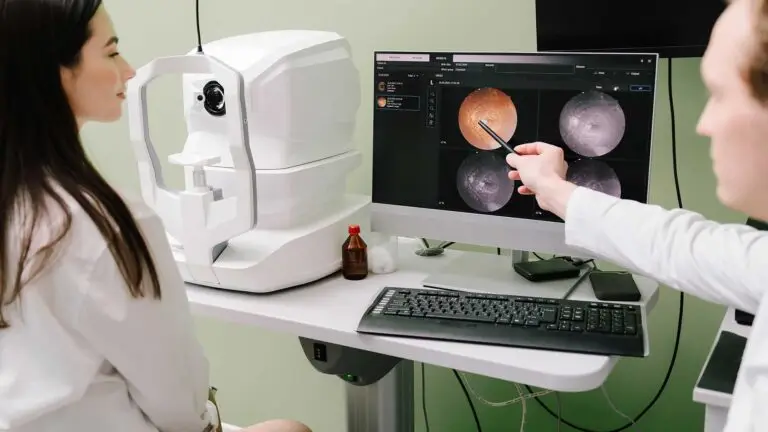
Optical coherence tomography (OCT) reveals detailed images of the retina, while artificial intelligence (AI) can help analyse them. Explore the potential benefits, current limitations and role of consultant oversight. Discover what these developments could mean for your next eye clinic assessment.
How Optical Coherence Tomography (OCT) Supports AI
OCT has become one of the most widely used imaging tests in medicine, producing detailed, digital images at enormous scale.¹
That combination, consistent images in very large numbers, is exactly what machine learning needs. Machine learning is a form of AI in which software learns patterns from examples. Researchers have trained systems to detect retinal disease and measure features such as fluid and atrophy, meaning thinning or loss of tissue, on OCT.²

What Artificial Intelligence (AI) Can Do
- Screening: an autonomous AI system, which produces a screening result without a clinician interpreting each image, analyses retinal photographs rather than OCT scans to detect diabetic retinopathy. This is damage to retinal blood vessels caused by diabetes. Following a pivotal trial, the system became the first of its kind to receive regulatory authorisation in the United States.³
- Measuring disease: AI can measure fluid and retinal thinning in wet and dry age-related macular degeneration (AMD) more consistently than the human eye, supporting decisions about treatment. AMD affects the central part of the retina used for detailed vision.²
- Prioritising referrals: by flagging urgent findings, AI can help direct patients who need prompt treatment to the front of the queue. An OCT study demonstrated that a trained system could make referral recommendations comparable to those of experts across several retinal diseases.⁴
The NHS diabetic eye screening programme uses retinal photographs to check for damage caused by diabetes. Trained staff assess these images and refer patients for further investigation or treatment where needed.⁵

What AI Cannot Do
AI systems are only as good as the data they are trained on and may perform less well in populations or on machines they have not seen. Their outputs can be wrong or unreliable, so appropriate checks remain essential.⁴ ⁵
An OCT-analysis tool does not replace a full clinical assessment. A clinician still needs to consider symptoms, examination findings, the patient’s priorities and the wider context before making an accountable treatment decision.
Some systems are designed to give a screening result without a clinician interpreting each image. At Blue Fin Vision®, our policy is that every scan and every AI output is reviewed by a consultant, who takes responsibility for the decision.

What This Means for Patients Now
- Faster answers: automated measurement of fluid and retinal thickness can make scan review quicker and more consistent.²
- Earlier warnings: AI-supported screening can flag people who need specialist review sooner.³
- Better monitoring: comparing scans over time is where computers excel, helping to detect subtle change in conditions such as wet AMD.²
- The same need for a consultant: diagnosis, treatment and the conversation about what matters to you remain clinical responsibilities.
Frequently Asked Questions
Will AI replace my eye doctor?
No. AI is a tool that supports clinicians, much as OCT itself did. At Blue Fin Vision®, diagnosis and treatment decisions always rest with your consultant.
Is my scan data used to train AI?
Only with appropriate legal safeguards and approvals. You can ask any clinic how your data is used and stored.
Can AI detect eye disease at home?
Home-monitoring devices are being developed and evaluated, but for now, symptoms and changes need assessment in clinic.
OCT Assessment at Our Eye Clinic
At our eye clinic, consultants use OCT imaging to assess the retina and explain the findings. We use modern imaging, including OCT, in every medical retina assessment, which focuses on conditions affecting the retina.
We also use AI-informed tools where they add accuracy, such as in surgical planning. Every scan is reviewed and every decision is made by a consultant.
When imaging shows wet AMD or macular oedema, swelling caused by fluid in the central retina, treatment can begin at our Harley Street clinic the same day or the following day. Our article on Macular Degeneration explains how the service works.
Read more about the scans and our service:
Discuss Your Eye Health With Blue Fin Vision®
Book a paid, consultant-led consultation with Blue Fin Vision® to discuss your eye health and whether OCT assessment is appropriate.
Consultations are available across London, Hertfordshire and Essex. Your consultant will explain which investigations are suitable and what the findings mean for your care. AI analysis is not automatically included in every assessment.
References
- Fujimoto J, Swanson E. The development, commercialization, and impact of optical coherence tomography. Invest Ophthalmol Vis Sci. 2016;57(9):OCT1-OCT13.
- Schmidt-Erfurth U, Sadeghipour A, Gerendas BS, Waldstein SM, Bogunović H. Artificial intelligence in retina. Prog Retin Eye Res. 2018;67:1-29.
- Abràmoff MD, Lavin PT, Birch M, Shah N, Folk JC. Pivotal trial of an autonomous AI-based diagnostic system for detection of diabetic retinopathy in primary care offices. NPJ Digit Med. 2018;1:39.
- De Fauw J, Ledsam JR, Romera-Paredes B, et al. Clinically applicable deep learning for diagnosis and referral in retinal disease. Nat Med. 2018;24:1342-1350.
- Syed O. UK NSC consults on use of artificial intelligence in the diabetic eye screening programme [Internet]. UK National Screening Committee; 2026 May 11 [cited 2026 Nov 2].
ABOUT THE AUTHOR
Mr Mfazo Hove
MBChB MD FRCOphth CertLRS
Consultant Ophthalmic Surgeon | Founder and Medical Director, Blue Fin Vision®
Mr Mfazo Hove is a Consultant Ophthalmic Surgeon with more than two decades of experience across NHS and private practice and more than 57,000 ophthalmic procedures performed. He trained for six and a half years at Moorfields Eye Hospital and served for five years as an NHS consultant at the Western Eye Hospital, Imperial College Healthcare NHS Trust.
He specialises in cataract surgery, refractive lens replacement, laser vision correction and implantable Collamer lenses (ICL). He is the founder and Medical Director of Blue Fin Vision®, a private ophthalmology group providing consultant-delivered care across London, Hertfordshire and Essex. His cataract surgery outcomes are publicly reported through the National Ophthalmology Database.
A ZEISS Key Opinion Leader and international speaker, Mr Hove is a contributing author to The Ophthalmologist and Ophthalmology Times. His writing examines how clinical governance and the organisation of ophthalmic services shape patients’ experiences and outcomes, with a focus on transparent outcome reporting, surgical training and long-term accountability for patient care.