



- Medically Reviewed by: Mr Mfazo Hove, Consultant Ophthalmic Surgeon
- Author: Mr Mfazo Hove
- Published: November 7, 2024
- Last Updated: October 29, 2026
TL;DR: Optical coherence tomography (OCT) is a quick, painless scan that uses light to create detailed cross-sectional images of the eye, a little like an ultrasound but with light instead of sound. It shows the individual layers of the retina, the light-sensitive tissue at the back of the eye, measures their thickness to thousandths of a millimetre, and reveals fluid, swelling or damage long before you might notice a change in your vision. It is now central to diagnosing and monitoring most retinal conditions.
An OCT scan shows what an eye examination can only suspect.
How Optical Coherence Tomography Works
OCT was first described in 1991.¹ It measures how light reflects back from the different layers of the eye and builds these reflections into a detailed image. Since then it has become one of the most widely used imaging tests in medicine.²
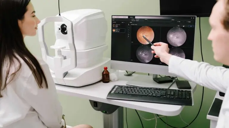
The scan is non-contact: you rest your chin, look at a target light, and the scan takes seconds.
What an OCT Scan Shows
- Fluid and swelling at the macula, the central part of the retina responsible for detailed vision. These changes can occur in wet age-related macular degeneration (AMD), where abnormal blood vessels leak; diabetic macular oedema, swelling caused by diabetes; retinal vein occlusion, a blockage in a vein draining the retina; and cystoid macular oedema, swelling with small fluid-filled spaces.
- Macular holes, gaps in the central retina, and membranes, thin layers of tissue that form on the retinal surface.
- Thinning of the retina in dry AMD, which causes gradual damage to the macula, and geographic atrophy, an advanced form involving loss of retinal cells.
The optic nerve, which carries visual signals to the brain, and the nerve fibre layer, the layer containing fibres from retinal nerve cells. These are assessed to help diagnose and monitor glaucoma, a condition that damages the optic nerve.

Why It Matters for Treatment
In wet AMD, OCT features such as fluid within or beneath the retina guide decisions about when to treat and when a treatment interval can be extended.³ In major treatment trials, OCT findings helped determine when repeat injections were needed.⁴
Monitoring with OCT means treatment is given when the eye needs it, not simply on a fixed timetable.
Types of OCT
- Posterior segment OCT images the back of the eye, including the retina, macula and optic nerve. Our article on posterior segment OCT explains it.
- OCT angiography maps blood flow in the retinal vessels without dye.⁵ Read about angio OCT.
- Anterior segment OCT images the front of the eye, for example to plan cataract surgery or implantable Collamer lens (ICL) surgery, where an additional lens is placed inside the eye to correct vision.
Is an OCT Safe?
Yes. OCT uses harmless near-infrared light, involves no radiation and does not touch the eye.
Your pupils may be dilated, or widened, with drops for the clearest images. This can blur your vision for a few hours, so avoid driving until it clears.
Preparing for Your Eye Clinic Visit
No special preparation is needed for your eye clinic appointment.
- Bring your glasses, a list of your medicines and any previous scans or clinic letters.
- Contact lenses can usually stay in for the scan itself, but you may be asked to remove them for other parts of the examination.
- If dilating drops are used, bring sunglasses and arrange not to drive for a few hours.
Understanding Your Results
Your consultant will usually show you the images. You are likely to see:
- A cross-section of the retina, a view through its layers, which appear as bands of different brightness. Fluid appears as dark spaces within or beneath these layers.
- A thickness map, a coloured circle showing where the macula is thicker or thinner than expected.
- A comparison with previous scans, showing whether swelling is improving or returning.
Normal values vary between machines, so scans are best compared on the same device over time.

How an OCT Scan Complements Your Eye Test
An OCT scan complements your eye test by providing detailed images of the layers beneath the retinal surface.
Test | What It Shows | What It Involves |
|---|---|---|
OCT | Layers of the retina, fluid and thickness | Seconds, non-contact |
Retinal photographs | The surface of the retina, haemorrhages (areas of bleeding) and new vessels | Seconds, a camera flash |
Fluorescein angiography, a dye test that photographs circulation in the eye | Blood flow and leakage | Dye injected into a vein in the arm |
Visual field test | How much side vision you have | Pressing a button when you see lights |
OCT is usually combined with at least one of these, because each answers a different question.²
OCT for Glaucoma
OCT also measures the thickness of the nerve fibre layer around the optic nerve. Thinning can appear before any loss of side vision is detectable, which helps diagnose glaucoma early and track whether treatment is working.
Frequently Asked Questions
Does OCT hurt?
No. Nothing touches your eye; you simply look at a light.
Does OCT use radiation?
No. OCT uses harmless near-infrared light.
My optometrist already did an OCT. Why repeat it?
High-street OCT is valuable for detecting problems. In clinic, scans are repeated on the same machine over time so that small changes can be measured precisely and treatment decisions made.
How often will I need scans?
It depends on the condition. During anti-vascular endothelial growth factor (anti-VEGF) treatment, which uses medicines to reduce abnormal blood vessel growth and leakage, OCT is usually done at every visit. For stable conditions, scans may be months apart.
Can OCT see problems outside the eye?
OCT can show swelling of the optic nerve, which may point to conditions elsewhere, and your consultant will arrange further investigation if needed.
At Blue Fin Vision®
OCT imaging is part of every medical retina assessment, an assessment of conditions affecting the retina. It is repeated at treatment visits to guide decisions.
Our clinics include:
- ZEISS CIRRUS 6000 OCT at Harley Street.
- Heidelberg SPECTRALIS imaging at Phoenix Hospital Chelmsford.
- REVO OCT at Chase Lodge Hospital and One Hatfield Hospital.
OCT scan pricing is set out on our private intravitreal injections page.
For more information about our care and preparing for treatment:
- Read about our medical retina service.
- Explore our patient information.
Book a consultation with Blue Fin Vision® to discuss your eye health and whether an OCT scan is appropriate.
References
- Huang D, Swanson EA, Lin CP, Schuman JS, Stinson WG, Chang W, Hee MR, Flotte T, Gregory K, Puliafito CA, Fujimoto JG. Optical coherence tomography. Science. 1991;254(5035):1178-81.
- Fujimoto J, Swanson E. The development, commercialization, and impact of optical coherence tomography. Invest Ophthalmol Vis Sci. 2016;57(9):OCT1-OCT13.
- Schmidt-Erfurth U, Waldstein SM. A paradigm shift in imaging biomarkers in neovascular age-related macular degeneration. Prog Retin Eye Res. 2016;50:1-24.
- Martin DF, Maguire MG, Ying GS, Grunwald JE, Fine SL, Jaffe GJ; Comparison of Age-related Macular Degeneration Treatments Trials (CATT) Research Group. Ranibizumab and bevacizumab for neovascular age-related macular degeneration. N Engl J Med. 2011;364(20):1897-908.
- Spaide RF, Fujimoto JG, Waheed NK, Sadda SR, Staurenghi G. Optical coherence tomography angiography. Prog Retin Eye Res. 2018;64:1-55.
ABOUT THE AUTHOR
Mr Mfazo Hove
MBChB MD FRCOphth CertLRS
Consultant Ophthalmic Surgeon | Founder and Medical Director, Blue Fin Vision®
Mr Mfazo Hove is a Consultant Ophthalmic Surgeon with more than two decades of experience across NHS and private practice and more than 57,000 ophthalmic procedures performed. He trained for six and a half years at Moorfields Eye Hospital and served for five years as an NHS consultant at the Western Eye Hospital, Imperial College Healthcare NHS Trust.
He specialises in cataract surgery, refractive lens replacement, laser vision correction and implantable Collamer lenses (ICL). He is the founder and Medical Director of Blue Fin Vision®, a private ophthalmology group providing consultant-delivered care across London, Hertfordshire and Essex. His cataract surgery outcomes are publicly reported through the National Ophthalmology Database.
A ZEISS Key Opinion Leader and international speaker, Mr Hove is a contributing author to The Ophthalmologist and Ophthalmology Times. His writing examines how clinical governance and the organisation of ophthalmic services shape patients’ experiences and outcomes, with a focus on transparent outcome reporting, surgical training and long-term accountability for patient care.