




- Medically Reviewed by: Mr Mfazo Hove, Consultant Ophthalmic Surgeon
- Author: Mr Mfazo Hove
- Published: December 19, 2024
- Last Updated: October 8, 2026
Understanding Corneal Decompensation
TL;DR: Corneal decompensation after a well-placed modern implantable collamer lens (ICL) is rare.¹ ² It happens when the endothelium, the cells lining the back of the cornea, becomes too depleted to keep this clear front surface of the eye transparent. Those cells do not regenerate, so the protection is measurement before ICL surgery and repeated counts throughout the life of the implant, so that any unexpected loss is identified before symptoms develop.
The endothelium cannot be felt, only measured. So we measure it.
Understanding Corneal Decompensation
The endothelium is a single layer of cells on the inner surface of the cornea. It pumps fluid out of the cornea to keep it thin and transparent. Everyone loses some of these cells slowly with age.
If the count falls far below normal, the cornea swells and vision becomes cloudy. That is corneal decompensation.
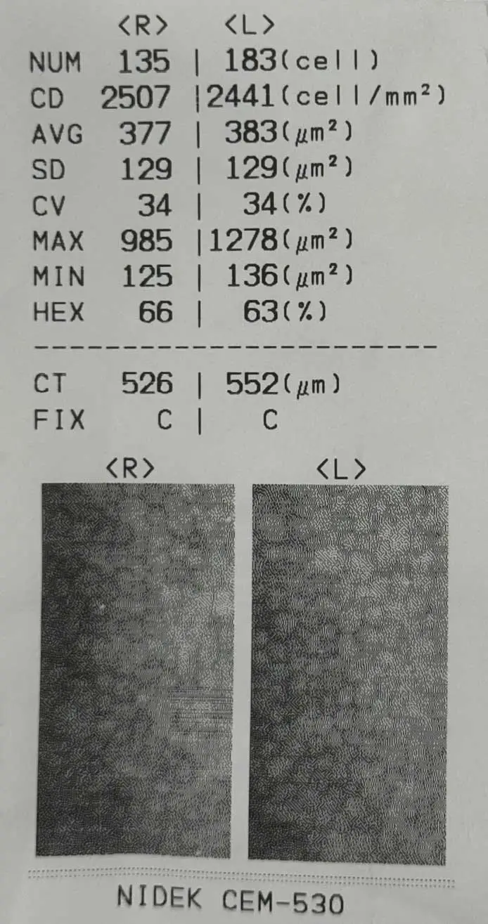
A specular microscopy report, produced using a microscope that photographs and analyses the cells lining the back of the cornea. Endothelial cell density, the number of cells in a measured area, is 2,507 cells per square millimetre in the right eye and 2,441 in the left. These figures are comfortably above the 2,000 threshold at which the Lifetime Monitoring Protocol™ triggers a consultant review.
The report also records how much cell size varies and the proportion of healthy six-sided cells.
Being above this review threshold does not by itself establish suitability for an ICL. That also depends on age, the depth of the space at the front of the eye and the criteria for the specific lens.
What Research Shows After ICL Surgery
Research has examined endothelial cell density and other outcomes after ICL surgery:
- Central-port ICLs, which have a small opening to allow fluid to flow through the lens, have shown endothelial cell density comparable with earlier designs, with no additional loss attributable to the central port.³
- Eight-year follow-up of central-port lenses confirmed long-term safety, with stable vision and prescription results.⁴
- In a 10-year study, endothelial cell density was tracked alongside vision, eye pressure and lens clarity.⁵
- Reviews of central-port lenses, one covering more than 2,900 eyes, compared endothelial cell density across studies and reported low rates of complications.¹ ²
Endothelial cell loss is recognised as a potential complication of any lens inside the eye.⁶
The risk rises if:
- The ICL is poorly sized.
- The space in front of the lens is too shallow.
- There is chronic inflammation, meaning persistent inflammation inside the eye.² ⁶
That is why suitability assessment measures the anterior chamber, the fluid-filled space at the front of the eye, and why sizing matters.

How Blue Fin Vision® Eye Clinic Monitors Your Cornea
Under the ICL Lifetime Monitoring Protocol™, our eye clinic measures your endothelial cell count by specular microscopy at our Harley Street centre.
The monitoring schedule includes:
- A count before surgery to establish your baseline, the starting measurement used for future comparisons.
- A count every year for as long as the ICL remains in your eye.
- More frequent checks if cells are being lost faster than expected.
Consultant review is triggered, at no additional cost, if your count trends below 2,000 cells per square millimetre or falls faster than the expected age-adjusted rate, meaning the loss normally expected at your age.
Options then include:
- Closer monitoring
- ICL exchange, replacing the existing implant with another lens
- ICL removal
These decisions are made with your full record on a single timeline.
If you develop increasing pain, light sensitivity, redness or worsening blurred vision, contact your surgeon or an eye casualty service without delay. Do not wait for your next scheduled monitoring appointment.⁷
Why Your Own Baseline Matters
A single count tells you where your cornea is. A series of counts tells you where it is heading.
- A result comfortably within the normal range can still warrant attention if it has fallen faster than expected since your previous measurements.
- A lower count that has been stable for years may need nothing more than continued monitoring.
That is why every measurement is compared with your own record, not only with a population reference range, the values typically found in a wider group of people.
The dataset belongs to you, and is available on request.
Further Reading
Read these supporting resources before your consultation:
- Our ICL suitability page.
- The phakic intraocular lens information, covering lenses implanted while your natural lens remains in place.
- Our ICL surgery checklist, which asks every provider how endothelial cells are tracked.
References
- Montés-Micó R, Ruiz-Mesa R, Rodríguez-Prats JL, Tañá-Rivero P. Posterior-chamber phakic implantable collamer lenses with a central port: a review. Acta Ophthalmol. 2021;99(3):e288-e301. doi:10.1111/aos.14599.
- Packer M. The Implantable Collamer Lens with a central port: review of the literature. Clin Ophthalmol. 2018;12:2427-38. doi:10.2147/OPTH.S188785.
- Goukon H, Kamiya K, Shimizu K, Igarashi A. Comparison of corneal endothelial cell density and morphology after posterior chamber phakic intraocular lens implantation with and without a central hole. Br J Ophthalmol. 2017;101(11):1461-5.
- Kamiya K, Shimizu K, Takahashi M, Ando W, Hayakawa H, Shoji N. Eight-year outcomes of implantation of posterior chamber phakic intraocular lens with a central port for moderate to high ametropia. Front Med (Lausanne). 2021;8:799078. doi:10.3389/fmed.2021.799078. PMID:34977099.
- Choi JH, Lim DH, Nam SW, Yang CM, Chung ES, Chung TY. Ten-year clinical outcomes after implantation of a posterior chamber phakic intraocular lens for myopia. J Cataract Refract Surg. 2019;45(11):1555-61. doi:10.1016/j.jcrs.2019.06.015. PMID:31587936.
- Fernandes P, González-Méijome JM, Madrid-Costa D, Ferrer-Blasco T, Jorge J, Montés-Micó R. Implantable collamer posterior chamber intraocular lenses: a review of potential complications. J Refract Surg. 2011;27(10):765-76. doi:10.3928/1081597X-20110617-01.
- Royal College of Ophthalmologists. Phakic intraocular lens implantation: patient information. London: Royal College of Ophthalmologists.
ABOUT THE AUTHOR
Mr Mfazo Hove
Consultant Ophthalmic Surgeon
MBChB MD FRCOphth CertLRS
Mr Mfazo Hove is a Consultant Ophthalmic Surgeon with experience spanning more than 57,000 procedures. He completed 6.5 years of specialist training at Moorfields Eye Hospital and served for five years as a consultant at the Western Eye Hospital, Imperial College Healthcare NHS Trust. He is the founder of Blue Fin Vision®, a consultant-led private ophthalmology practice operating across London, Essex, and Hertfordshire. His clinical expertise encompasses advanced cataract surgery, refractive lens replacement, laser vision correction, and implantable Collamer lenses (ICL).
A ZEISS Key Opinion Leader, Mr Hove is a respected international speaker with five invited engagements across seven cities in 2026:
- ZEISS China tour (Changsha, Shanghai, and Hangzhou, April – ZEISS APAC User Meeting)
- RCOphth Annual Congress – May – Manchester
- ZEISS EMEA User Meeting (Istanbul)
- ZEISS Lausanne User Meeting (Lausanne)
- European Society of Cataract and Refractive Surgeons Annual Congress (ESCRS, London)
Schedule Your Consultation Today
Book a consultation with Blue Fin Vision® in London, Hertfordshire or Essex to discuss ICL surgery, corneal decompensation risk and your long-term monitoring needs.


