




- Medically Reviewed by: Mr Mfazo Hove, Consultant Ophthalmic Surgeon
- Author: Mr Mfazo Hove
- Published: February 10, 2025
- Last Updated: October 20, 2026
TL;DR: A pinguecula is a small, raised, yellowish patch on the conjunctiva, the thin clear tissue covering the white of the eye. It is benign, meaning non-cancerous, and usually appears beside the cornea, the clear front surface of the eye, nearest the nose. Most need only lubricating drops and sun protection, although removal may be considered for persistent irritation, contact lens discomfort or cosmetic concerns.
Is Pinguecula Dangerous?
A pinguecula is harmless. Whether it is worth removing depends on how much it troubles you, not on any danger it poses.
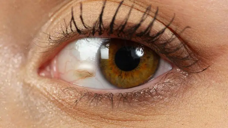
A pinguecula: the small yellowish patch on the white of the eye, beside the cornea on the side nearest the nose.
How Common Is Pinguecula?
Very. In a study of nearly a thousand adults aged 50 and over in a rural community near Shanghai, three quarters had a pinguecula.¹
Figures vary between populations, but pinguecula is one of the most common changes seen on the surface of the eye. In the Blue Mountains Eye Study in Australia, pinguecula was far more common than pterygium.²
Pinguecula Causes and Risk Factors
Pinguecula is linked to sunlight, wind and age. In the Shanghai study, increasing age and more than two hours of daily sunlight were the strongest associations.¹
Pinguecula Symptoms
Many people never notice theirs. Common pinguecula symptoms include:
- A visible yellowish bump on the white of the eye.
- Grittiness, dryness or a feeling that something is in the eye.
- Episodes of redness and inflammation, called pingueculitis.
- Discomfort with contact lenses.
Inflamed pingueculae are associated with damage to the surface of the nearby cornea and a less stable tear film, the thin layer of tears covering the eye. In one study, 72% of eyes with an inflamed pinguecula showed damage to the corneal surface next to it.³
A raised pinguecula can disrupt the way tears spread across the eye, and removing it can resolve dry eye symptoms that had not responded to drops.⁴
Is It the Same as a Pterygium?
No. A pinguecula stays on the white of the eye. A pterygium is a fleshy, wedge-shaped growth that extends onto the cornea itself and can affect vision. Our article on pinguecula and pterygium explains the difference.
Pinguecula Treatment
Pinguecula treatment depends on how much it troubles you. When one becomes persistently irritated, interferes with contact lenses or bothers you cosmetically, it can be removed.
Without Surgery
Lubricating drops, sunglasses and short courses of anti-inflammatory drops during flare-ups control symptoms for most people.
Surgical Excision
Surgical excision means surgically removing the pinguecula. It is performed under local anaesthetic, which numbs the treatment area, in a short procedure aiming to remove the raised tissue in a single sitting. At Blue Fin Vision®, surgical excision is now the treatment we recommend for most patients.
Blue Fin Vision® has removed more than 100 pingueculae between October 2025 and September 2026.
Argon Laser
Laser can flatten a pinguecula without an incision. A small published study reported good cosmetic results with laser in selected pingueculae.⁵
In our own experience, a meaningful proportion of patients needed repeat laser treatment, which is why excision is now our usual treatment, even though many patients arrive hoping for laser as the gentler option. Our article on pinguecula removal in London explains why.
Laser remains available at Harley Street for selected small pingueculae.
At Blue Fin Vision®
Surgical removal of pinguecula is available at Harley Street in London, Phoenix Hospital Chelmsford and One Hatfield Hospital. Treatment is priced per pinguecula, as shown on our costs page.
Before your consultation, read these supporting resources:
- Our pterygium and pinguecula removal page.
- Our suitability page.
- Our risks page.
- Our patient information.
Book a consultation at a Blue Fin Vision® eye clinic to discuss your pinguecula treatment options.
References
- Le Q, Xiang J, Cui X, Zhou X, Xu J. Prevalence and associated factors of pinguecula in a rural population in Shanghai, Eastern China. Ophthalmic Epidemiol. 2015;22(2):130-8.
- Panchapakesan J, Hourihan F, Mitchell P. Prevalence of pterygium and pinguecula: the Blue Mountains Eye Study. Aust N Z J Ophthalmol. 1998;26 Suppl 1:S2-5.
- Küçük E, Yilmaz U, Zor KR. Corneal epithelial damage and impaired tear functions in patients with inflamed pinguecula. J Ophthalmol. 2018;2018:2474173. doi:10.1155/2018/2474173.
- Napoli PE, Sanna R, Iovino C, Fossarello M. Resolution of pinguecula-related dry eye disease after argon laser photocoagulation. Int Med Case Rep J. 2017;10:247-50.
- Ahn SJ, Shin KH, Kim MK, Wee WR, Kwon JW. One-year outcome of argon laser photocoagulation of pinguecula. Cornea. 2013;32(7):971-5.
ABOUT THE AUTHOR
Mr Mfazo Hove
MBChB MD FRCOphth CertLRS
Consultant Ophthalmic Surgeon | Founder and Medical Director, Blue Fin Vision®
Mr Mfazo Hove is a Consultant Ophthalmic Surgeon with more than two decades of experience across NHS and private practice and more than 57,000 ophthalmic procedures performed. He trained for six and a half years at Moorfields Eye Hospital and served for five years as an NHS consultant at the Western Eye Hospital, Imperial College Healthcare NHS Trust.
He specialises in cataract surgery, refractive lens replacement, laser vision correction and implantable Collamer lenses (ICL). He is the founder and Medical Director of Blue Fin Vision®, a private ophthalmology group providing consultant-delivered care across London, Hertfordshire and Essex. His cataract surgery outcomes are publicly reported through the National Ophthalmology Database.
A ZEISS Key Opinion Leader and international speaker, Mr Hove is a contributing author to The Ophthalmologist. His writing examines how clinical governance and the organisation of ophthalmic services shape patients’ experiences and outcomes, with a focus on transparent outcome reporting, surgical training and long-term accountability for patient care.