




- Medically Reviewed by: Mr Mfazo Hove, Consultant Ophthalmic Surgeon
- Author: Mr Mfazo Hove
- Published: February 11, 2025
- Last Updated: October 20, 2026
TL;DR: The main difference between pinguecula and pterygium is where they grow: a pinguecula stays on the white of the eye, while a pterygium extends onto the cornea, the clear front surface of the eye. Both are benign, meaning non-cancerous, growths of the conjunctiva, the thin clear tissue covering the white of the eye. A pterygium can change the cornea’s shape and affect vision, which influences treatment and how closely it needs monitoring.
What are Pinguecula and Pterygium?
A pinguecula is a benign, yellowish raised bump that forms on the conjunctiva, the clear membrane covering the white part of the eye (sclera). It typically appears on the nasal side of the eye and does not grow onto the cornea.
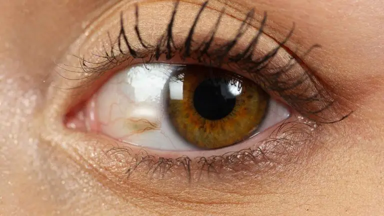
A pterygium, often referred to as “surfer’s eye”, is a wedge-shaped growth of fleshy tissue that starts on the conjunctiva and can extend onto the cornea. It’s usually pink or reddish in colour and has a more triangular shape compared to a pinguecula.

What Is the Difference Between Pinguecula and Pterygium?
Feature | Pinguecula | Pterygium |
|---|---|---|
Appearance | Yellowish, raised and rounded | Pink or red, fleshy and wedge-shaped |
Where It Sits | On the white of the eye, beside the cornea | Extends from the white of the eye onto the cornea |
Effect on Vision | Does not grow onto the cornea. An inflamed pinguecula can disturb the tear film, the thin layer of tears covering the eye, and affect comfort and visual quality. | Can cause astigmatism, an uneven curvature of the cornea that can blur vision, or reach the line of sight if large. |
How Common | Very common | Less common |
After Removal | Recurrence, meaning the growth returning, is uncommon. | Recurrence is the main concern. |
Blue Fin Vision® Treatment | Surgical removal for most; laser for selected small growths. | Removal with a conjunctival autograft, a graft of your own healthy conjunctival tissue. |
In the Blue Mountains Eye Study, pinguecula was considerably more common than pterygium.¹ The prevalence of pterygium varies widely between countries and climates, and rises with outdoor exposure.²
Why They Form
Both are linked to sunlight, wind and dust, and are associated with long-term exposure to ultraviolet light from the sun. Light striking the side of the eye can be focused by the cornea onto the inner corner, which may explain why both growths usually appear on the side nearest the nose.³
In a population study of pinguecula, age and more than two hours of daily sunlight were the strongest associations.⁴
How Each Is Treated
Pinguecula Treatment
Lubricating drops and sun protection come first.
If a pinguecula remains irritated or bothers you cosmetically, surgical excision, meaning surgical removal, aims to remove it in one procedure.
Laser can work for small growths,⁵ but in our experience a meaningful proportion of patients needed repeat treatment, so excision is now our usual treatment.
Pterygium Treatment
Surgery is considered when a pterygium causes persistent symptoms, is growing towards the centre of the cornea or is affecting vision.
The pterygium is removed, and the bare area is covered with a thin graft of your own healthy conjunctiva from under the upper eyelid. This is called a conjunctival autograft.
A Cochrane review found that conjunctival autograft carried a lower risk of recurrence than amniotic membrane grafting, which uses donated tissue from the membrane surrounding a baby during pregnancy.⁶
Recovery at a Glance
After Pinguecula Removal
- Redness: one to three weeks.
- Return to work: most people return within two to five days.
After Pterygium Removal With an Autograft
- Redness: two to six weeks.
- Return to work: most people return within three to seven days.
- Full healing: three to six months.
At Blue Fin Vision®
Pinguecula surgery is available at Harley Street in London, Phoenix Hospital Chelmsford and One Hatfield Hospital.
Pterygium surgery is available at Weymouth Street Hospital in London, Chelmsford and Hatfield.
Read these supporting resources before your consultation:
- Our pterygium and pinguecula removal page.
- Our frequently asked questions (FAQs).
- Our costs page.
- Our patient information.
Book a consultation at a Blue Fin Vision® eye clinic to discuss your pinguecula or pterygium treatment options.
References
- Panchapakesan J, Hourihan F, Mitchell P. Prevalence of pterygium and pinguecula: the Blue Mountains Eye Study. Aust N Z J Ophthalmol. 1998;26 Suppl 1:S2-5.
- Liu L, Wu J, Geng J, Yuan Z, Huang D. Geographical prevalence and risk factors for pterygium: a systematic review and meta-analysis. BMJ Open. 2013;3(11):e003787. doi:10.1136/bmjopen-2013-003787.
- Coroneo MT. Pterygium as an early indicator of ultraviolet insolation: a hypothesis. Br J Ophthalmol. 1993;77(11):734-9.
- Le Q, Xiang J, Cui X, Zhou X, Xu J. Prevalence and associated factors of pinguecula in a rural population in Shanghai, Eastern China. Ophthalmic Epidemiol. 2015;22(2):130-8.
- Ahn SJ, Shin KH, Kim MK, Wee WR, Kwon JW. One-year outcome of argon laser photocoagulation of pinguecula. Cornea. 2013;32(7):971-5.
- Clearfield E, Muthappan V, Wang X, Kuo IC. Conjunctival autograft for pterygium. Cochrane Database Syst Rev. 2016;2(2):CD011349. doi:10.1002/14651858.CD011349.pub2.
ABOUT THE AUTHOR
Mr Mfazo Hove
MBChB MD FRCOphth CertLRS
Consultant Ophthalmic Surgeon | Founder and Medical Director, Blue Fin Vision®
Mr Mfazo Hove is a Consultant Ophthalmic Surgeon with more than two decades of experience across NHS and private practice and more than 57,000 ophthalmic procedures performed. He trained for six and a half years at Moorfields Eye Hospital and served for five years as an NHS consultant at the Western Eye Hospital, Imperial College Healthcare NHS Trust.
He specialises in cataract surgery, refractive lens replacement, laser vision correction and implantable Collamer lenses (ICL). He is the founder and Medical Director of Blue Fin Vision®, a private ophthalmology group providing consultant-delivered care across London, Hertfordshire and Essex. His cataract surgery outcomes are publicly reported through the National Ophthalmology Database.
A ZEISS Key Opinion Leader and international speaker, Mr Hove is a contributing author to The Ophthalmologist and Ophthalmology Times. His writing examines how clinical governance and the organisation of ophthalmic services shape patients’ experiences and outcomes, with a focus on transparent outcome reporting, surgical training and long-term accountability for patient care.


