



- Medically Reviewed by: Mr Mfazo Hove, Consultant Ophthalmic Surgeon
- Author: Mr Mfazo Hove
- Published: November 20, 2024
- Last Updated: November 5, 2026
TL;DR: Diabetic retinopathy is damage caused by diabetes to the small blood vessels of the retina, the light-sensitive layer at the back of the eye, and its early stages cause no symptoms. As it progresses, vessels can leak and cause swelling at the macula, the central area responsible for detailed vision, or close off and trigger fragile new vessels that bleed. Both complications are treatable, and good control of blood sugar, blood pressure and cholesterol reduces the risk of either happening.
Diabetic retinopathy rarely announces itself. Screening finds it before you do.

What Causes Diabetic Retinopathy?
Raised blood sugar over years damages the lining of the retinal capillaries, the smallest blood vessels in the retina. They leak, form small bulges called microaneurysms and, in some areas, close altogether. ¹ Retinopathy is usually described in two broad forms:
- Non-proliferative: leaking and damaged vessels, without new vessel growth.
- Proliferative: new, fragile vessels grow in response to areas starved of oxygen.
Diabetic maculopathy means diabetes-related damage affecting the macula. Diabetic macular oedema specifically means swelling caused by fluid leakage. This swelling can occur at any stage of retinopathy and is the most common cause of reduced vision.
Our articles explain these changes in more detail:
Why Screening Works
Diabetic retinopathy used to be the leading cause of certified blindness in working-age adults in England and Wales. It no longer is, a change attributed in part to national screening and earlier treatment. ²
The NHS Diabetic Eye Screening Programme photographs the retina of everyone with diabetes aged 12 and over at regular intervals, and refers people who need specialist care. ³
If you have diabetes, attend every screening appointment, even when your sight feels perfect.
Reducing Your Risk
Landmark trials showed that better blood sugar control in type 1 diabetes, and tighter blood pressure control in type 2 diabetes, substantially reduce the development and progression of retinopathy. ⁴ ⁵ Stopping smoking and managing cholesterol help too.
Retinopathy can progress faster in pregnancy, so extra checks are arranged.


Diabetic Retinopathy Treatment
Macular Oedema
Anti-vascular endothelial growth factor (anti-VEGF) injections reduce leakage from retinal blood vessels and help control abnormal vessel growth. They are the main treatment when swelling involves the centre of the macula, with the decision based on your vision and the full clinical findings, not the scan alone.
In a large comparative trial, all three medicines studied improved vision, and aflibercept gave the greatest gains when vision was poorer at the start. ⁶
Proliferative Retinopathy
Laser treatment to the peripheral retina, the outer part of the retina, reduces the risk of severe sight loss. Injections are sometimes used as well.
Bleeding or Scar Tissue
Vitrectomy surgery removes the vitreous gel inside the eye. It can clear blood from the eye or relieve traction, pulling caused by scar tissue, on the retina.
Who Is at Risk?
Everyone with diabetes, type 1 or type 2, is at risk. The risk is higher with: ¹
- A longer duration of diabetes.
- Higher average blood sugar, measured by HbA1c, a blood test reflecting average blood glucose over approximately two to three months.
- High blood pressure and raised cholesterol.
- Kidney disease.
- Pregnancy.
- Smoking.

Diabetic Retinopathy Symptoms
Early retinopathy causes no symptoms. When symptoms do occur, they include:
- Blurred or fluctuating vision, often from macular swelling.
- Difficulty reading or seeing fine detail.
- New floaters, small shapes drifting across your vision, or dark patches, which can mean bleeding inside the eye.
- Sudden loss of vision, which needs same-day assessment.
For sudden sight loss, seek immediate help through an urgent eye service or eye casualty. If you cannot access one, go to A&E. For sudden worsening or blurred vision, contact an eye clinic urgently, request an urgent GP appointment or contact NHS 111. Do not wait for your next screening appointment. ⁷

Understanding Ischaemic Retinopathy: The Wembley Stadium Analogy
The Eye as Wembley Stadium
Imagine your eye is like Wembley stadium, filled with fans (representing your retina) who can’t leave.
The Problem: Blocked Gates
In severe diabetic retinopathy or vein occlusion:
- Gates are blocked, preventing food from coming in and rubbish from going out
- This represents blocked blood vessels that normally:
- Bring in oxygenated blood to nourish the retina
- Remove waste products
The Consequences: Hungry Fans Cause Chaos
Without proper blood flow:
- Hungry fans (retinal cells) cause a ruckus
- They throw rubbish around (vitreous haemorrhage)
- Tear up seats and fight (retinal damage)
The Solution: Laser Treatment
Since we can’t unblock the gates:
- Laser treatment “wipes out” sections of fans
- This continues until remaining fans have enough food
- Treatment starts at the top of the stadium (cheap seats = peripheral retina)
- Repeated until enough fans are removed to calm the situation
The Result
- Remaining retina gets sufficient blood supply
- Stops producing vascular endothelial growth factor
- Prevents growth of new abnormal blood vessels (neovascularisation)
Important Note
The laser generally avoids the pitch (central retina):
- This is the most important part of the stadium
- In the eye, it’s responsible for detailed vision
Macular Oedema: A Waterlogged Pitch
- Macular oedema is like a waterlogged pitch in our stadium
- Laser treatment burns small bits of the pitch
- This helps to “dry out” the area (reduce the macular oedema)
What Happens at Your Eye Clinic Appointment
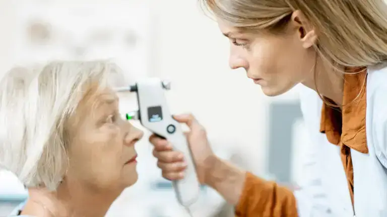
Your eye clinic appointment assesses your vision, retinal health and any need for treatment.
- Vision testing and eye pressure measurement.
- Dilating drops, which widen your pupils, and a full examination of the retina, including its outer edges.
- An optical coherence tomography (OCT) scan, which produces detailed cross-sectional images of the retina, to check the macula for swelling.
- Wide-field photographs, which capture a broad view of the retina, or angiography, imaging of the retinal blood vessels and circulation, where needed to map areas without a blood supply and any new vessels.
- A written plan covering monitoring, any treatment and advice for your GP.
Laser Treatment Explained
For proliferative retinopathy, laser is applied to the outer retina in one or more sessions. It is done in clinic with numbing drops and a contact lens on the eye.
Most people feel a series of bright flashes; some find it uncomfortable. The aim is to protect central vision by reducing the signal that encourages abnormal blood vessel growth.
Heavy treatment can reduce night vision and side vision, which can occasionally affect driving, and your consultant will discuss this.
Diabetes, Medicines and Your Eyes
A rapid, large improvement in blood sugar can occasionally cause a temporary worsening of retinopathy, so if your diabetes treatment changes significantly, tell your eye team. Our article on GLP-1 Medications and Your Vision discusses one example.
None of this is a reason to delay better diabetes control; it is a reason for closer eye monitoring.
A Prevention Checklist
- Attend every diabetic eye screening appointment.
- Know your HbA1c, blood pressure and cholesterol, and work with your GP to keep them on target. ⁴ ⁵
- Stop smoking.
- Tell your GP or eye team promptly about any change in your vision.
- If you are planning a pregnancy, ask about extra eye checks.
Frequently Asked Questions
My vision is perfect. Do I really need screening?
Yes. Retinopathy is often advanced before it affects vision, and treatment works best before sight is lost.
Will I go blind?
Most people with diabetes never lose significant vision. With screening and timely treatment, severe sight loss from diabetic retinopathy is much less common than it was. ²
Can retinopathy be reversed?
Early changes can stabilise or improve with better control of blood sugar and blood pressure. Treatment can reduce swelling and control new vessels, but vision already lost may not fully return.
Does laser damage my sight?
Laser is designed to protect central vision. It can reduce side and night vision, a trade-off that is worthwhile when proliferative disease threatens severe sight loss.
Is diabetic eye screening the same as an eye test?
No. Screening photographs the retina to look for retinopathy; it does not check whether you need glasses. You still need regular eye tests with your optometrist.
At Blue Fin Vision®
We assess retinopathy with OCT imaging and a wide examination of the retina.
Where diabetic macular oedema needs treatment, our service is built around rapid access. We arrange a consultation and OCT scan, typically the same day at your nearest suitable site, and an injection at our Harley Street clinic the same day or the following day. This is followed by an organised next step with us or through the NHS.
We write to your GP and diabetes team about blood sugar, blood pressure and cholesterol. Vitreoretinal surgery, surgery involving the retina and vitreous gel, is available within the same team through Professor Mahmut Dogramaci.
Read more about the condition and our services:
- Our article on Diabetic Maculopathy explains how the service works.
- Our Medical Retina page.
- Our Vitreoretinal Surgery page.
The Royal National Institute of Blind People (RNIB) and our patient resources provide further information:
- RNIB’s free Understanding Eye Conditions Related to Diabetes guide.
- Our Patient Information.
Book a consultation with Blue Fin Vision® to discuss your diabetic retinopathy assessment, treatment and monitoring, with consultant-led care at clinics across London, Hertfordshire and Essex.
References
- Cheung N, Mitchell P, Wong TY. Diabetic retinopathy. Lancet. 2010;376(9735):124-36.
- Liew G, Michaelides M, Bunce C. A comparison of the causes of blindness certifications in England and Wales in working age adults (16-64 years), 1999-2000 with 2009-2010. BMJ Open. 2014;4(2):e004015.
- Scanlon PH. The English National Screening Programme for diabetic retinopathy 2003-2016. Acta Diabetol. 2017;54(6):515-25.
- Diabetes Control and Complications Trial Research Group. The effect of intensive treatment of diabetes on the development and progression of long-term complications in insulin-dependent diabetes mellitus. N Engl J Med. 1993;329(14):977-86.
- UK Prospective Diabetes Study Group. Tight blood pressure control and risk of macrovascular and microvascular complications in type 2 diabetes: UKPDS 38. BMJ. 1998;317(7160):703-13.
- Diabetic Retinopathy Clinical Research Network; Wells JA, Glassman AR, Ayala AR, Jampol LM, Aiello LP, Antoszyk AN, et al. Aflibercept, bevacizumab, or ranibizumab for diabetic macular edema. N Engl J Med. 2015;372(13):1193-203.
- NHS. Diabetic retinopathy [Internet]. NHS; [cited 2026 Nov 4].
ABOUT THE AUTHOR
Mr Mfazo Hove
MBChB MD FRCOphth CertLRS
Consultant Ophthalmic Surgeon | Founder and Medical Director, Blue Fin Vision®
Mr Mfazo Hove is a Consultant Ophthalmic Surgeon with more than two decades of experience across NHS and private practice and more than 57,000 ophthalmic procedures performed. He trained for six and a half years at Moorfields Eye Hospital and served for five years as an NHS consultant at the Western Eye Hospital, Imperial College Healthcare NHS Trust.
He specialises in cataract surgery, refractive lens replacement, laser vision correction and implantable Collamer lenses (ICL). He is the founder and Medical Director of Blue Fin Vision®, a private ophthalmology group providing consultant-delivered care across London, Hertfordshire and Essex. His cataract surgery outcomes are publicly reported through the National Ophthalmology Database.
A ZEISS Key Opinion Leader and international speaker, Mr Hove is a contributing author to The Ophthalmologist and Ophthalmology Times. His writing examines how clinical governance and the organisation of ophthalmic services shape patients’ experiences and outcomes, with a focus on transparent outcome reporting, surgical training and long-term accountability for patient care.