


- Medically Reviewed by: Mr Mfazo Hove, Consultant Ophthalmic Surgeon
- Author: Mr Mfazo Hove
- Published: August 12, 2024
- Last Updated: September 11, 2026
TL;DR: Strip away the marketing and ask: what does laser eye surgery do? At a fundamental optical level, laser eye surgery does exactly one thing: it changes the shape of your cornea by microns so that light entering your eye lands in focus on the retina, although different procedures achieve this in different ways. This page explains the mechanism, treatment planning, tissue limits, durability and boundaries in plain English; for the broader introduction, procedures, assessment and surgery day, read What Is Laser Eye Surgery?.
How an Eye Focuses Before Laser
Light from the world enters through the cornea, which does most of the bending. It then passes through the pupil, is fine-tuned by the natural lens and lands, in a perfect eye, exactly on the retina.
Focus is therefore a matching problem. The combined power of the cornea and lens must match the length of the eye. A prescription is simply a measurement of the mismatch, and a dioptre is its unit. Conceptually, it describes how strong a correcting lens must be to move your focal point onto the retina.
Minus numbers mean the system is too powerful or the eye too long, while plus numbers mean the reverse. The cylinder number measures astigmatism, the meridian-to-meridian inconsistency. Laser eye surgery resolves the mismatch not by adding a lens but by editing the most powerful element in the system: the cornea itself.
What the Laser Changes in Myopia
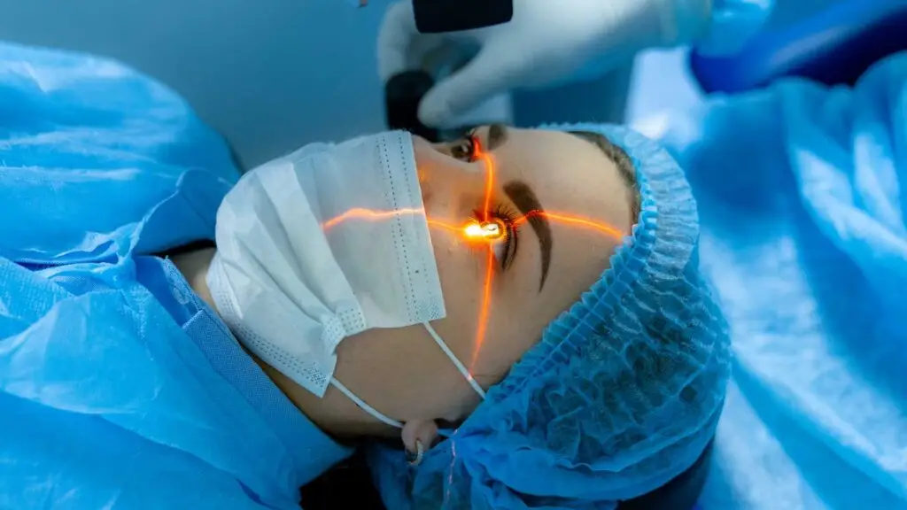
The explanation below primarily describes excimer-laser treatments, including LASIK and PRK. SMILE reshapes the cornea differently: a femtosecond laser creates a small disc of tissue, known as a lenticule, inside the cornea. The lenticule is then removed through a small incision to alter the corneal shape.⁵
In myopia, the focal point sits in front of the retina, so the cornea must become less powerful. The excimer laser flattens the central cornea by removing a precisely calculated, lens-shaped profile of tissue. The profile is deepest centrally and tapers outwards until the cornea’s curvature bends light less and the focal point slides back onto the retina.
The bigger the correction, the more tissue the profile requires. This is where the tissue budget below enters.
What the Laser Changes in Myopia
In hyperopia, the focal point sits behind the retina, so the cornea must become more powerful. You cannot add tissue with a laser that only removes it.
The solution is elegant. The treatment removes tissue in an annulus, a ring around the centre. This allows the central cornea, which is left relatively untouched, to stand proportionally steeper. It gains power and draws the focal point forward.
Hyperopic profiles are technically more demanding and range-limited compared with myopic ones. This is one reason high hyperopia often routes towards lens-based surgery instead.
What It Changes in Astigmatism
An astigmatic cornea carries two curvatures, steep in one meridian and flatter in the other. The treatment is therefore asymmetric by design. More tissue is removed along the steep meridian and less along the flat until the surface is regularised and light converges to a single point.
The correction is folded into the same profile as your sphere, creating one integrated reshaping. Its orientation matters absolutely, which is why alignment technology exists, as explained below.
The full astigmatism treatment, including the regular-versus-irregular distinction that decides safety, is covered in Will Laser Eye Surgery Fix Astigmatism?.

How Much Tissue, Across What Zone, And Why Your Pupil Matters
The quantities are microscopic. The excimer removes fractions of a micron per pulse, and a whole correction is written in a layer thinner than a human hair.
The reshaping is applied across an optical zone, the central disc of the cornea sculpted to the new curve. This is blended outwards through a transition zone. The optical zone is planned with your pupil in mind.
In dim light, the pupil widens. If it opens beyond the well-corrected zone, light entering through the periphery is focused differently. This is one mechanistic contributor to night-time haloes and starbursts.
Larger corrections and smaller zones were prominent risk factors in the classic night-vision literature.⁶ This is precisely why zone planning is part of the physics of your treatment rather than a preference.
Wavefront, Topography Guidance and Eye Tracking
Three technologies refine where and how the profile is delivered.
Wavefront-guided treatment measures the eye’s entire optical fingerprint, including its higher-order imperfections beyond sphere and cylinder. It then shapes the profile to that information.
Topography-guided treatment builds the profile from the cornea’s own surface map. This can be valuable where surface irregularity is the story.
Eye tracking with cyclotorsion control solves the delivery problem. The tracker follows the eye’s position hundreds of times a second, while registration aligns the treatment with your eye’s landmarks. Rotational compensation matters because the eye rotates slightly between sitting and lying. Without compensation, this could twist an astigmatic axis.
None of these technologies changes what the laser does. They sharpen where it does it.

Why A Tissue Budget Exists
The cornea is structural as well as optical. It must stay strong for life, so every plan balances the tissue removed against the tissue retained. A correction that would overdraw the budget is declined.
This is not conservatism for its own sake. The rare but potentially serious late complication of corneal refractive surgery is ectasia, progressive weakening and distortion. Respecting the tissue budget, together with careful screening of the corneal shape, is designed to minimise that risk. However, no responsible surgeon should imply that biological risk can ever be reduced to zero.⁴
Can the Cornea Heal Back to Its Old Shape?
Essentially no, and this is the durability question answered mechanistically. The excimer removes tissue from the corneal stroma, a layer that does not regenerate removed volume. The reshaping is therefore structural and lasting.
What healing does do, especially after surface techniques, is remodel the outermost cell layer. In a minority of eyes, this contributes to regression, a small drift back towards the old prescription over time.
Regression is the exception, not the rule, in well-selected modern treatments.¹ ³ Where it produces a symptomatic residual error, the answer is enhancement rather than resignation.
What If the Correction Is Slightly Wrong?
Then it is finished, not failed. Biology delivers a distribution around every target, and a small proportion of eyes land slightly off.
A symptomatic residual error may be managed with refractive enhancement where this is clinically appropriate and the tissue budget allows. Provider policies vary. Enhancement may be included for a defined period, subject to eligibility, charged separately or handled under provider-specific terms. Ask to see the written policy before surgery.
At Blue Fin Vision®, enhancement after self-pay laser vision correction is fully covered within 24 months for eligible patients under the written Enhancement Policy.

What Laser Does Not Change
Your natural lens. The treatment lives entirely in the cornea. The lens continues its lifelong ageing untouched.
Accommodation. The young lens’s ability to refocus, and its gradual mid-40s loss, presbyopia, proceed on their own schedule. PRESBYOND® uses a blended-vision corneal profile to extend the range of clear vision. It manages the effects of presbyopia at the corneal level but does not reverse the ageing process within the natural lens.
Your retina. Nothing at the back of the eye is treated or altered.
Axial length. A myopic eye is optically corrected, not shortened. The eye’s anatomy behind the cornea is exactly as it was.
Future cataract. The lens that will one day cloud is the same lens, on the same timetable. A lasered cornea makes those eventual lens calculations more demanding, a point covered elsewhere on this site.
Underlying myopic retinal risk. The long myopic eye keeps its long-eye retinal considerations after perfect laser because the laser corrected the optics, not the anatomy. Perfect unaided vision is not a discharge from sensible retinal care.
So, What Does Laser Eye Surgery Do?
One thing, superbly, for the right eyes: it re-sculpts the cornea, micron by planned micron, so your own optics do the focusing your glasses used to, with the evidence base to match.² ³
Understanding the single mechanism explains both the operation’s strengths and its boundaries better than any brochure.
References
- Shortt AJ, Allan BD, Evans JR. Laser-assisted in-situ keratomileusis (LASIK) versus photorefractive keratectomy (PRK) for myopia. Cochrane Database Syst Rev. 2013;(1):CD005135. PMID: 23440799.
- Solomon KD, Fernández de Castro LE, Sandoval HP, Biber JM, Groat B, Neff KD, Ying MS, French JW, Donnenfeld ED, Lindstrom RL; Joint LASIK Study Task Force. LASIK world literature review: quality of life and patient satisfaction. Ophthalmology. 2009;116(4):691-701. PMID: 19344821.
- Sandoval HP, Donnenfeld ED, Kohnen T, Lindstrom RL, Potvin R, Tremblay DM, Solomon KD. Modern laser in situ keratomileusis outcomes. J Cataract Refract Surg. 2016;42(8):1224-1234. PMID: 27531300.
- Randleman JB, Woodward M, Lynn MJ, Stulting RD. Risk assessment for ectasia after corneal refractive surgery. Ophthalmology. 2008;115(1):37-50. PMID: 17624434.
- Reinstein DZ, Archer TJ, Gobbe M. Small incision lenticule extraction (SMILE) history, fundamentals of a new refractive surgery technique and clinical outcomes. Eye Vis (Lond). 2014;1:3. PMID: 26605350.
- Pop M, Payette Y. Risk factors for night vision complaints after LASIK for myopia. Ophthalmology. 2004;111(1):3-10. PMID: 14711706.
ABOUT THE AUTHOR
Mr Mfazo Hove
Consultant Ophthalmic Surgeon
MBChB MD FRCOphth CertLRS
Mr Mfazo Hove is a Consultant Ophthalmic Surgeon with experience spanning more than 57,000 procedures. He completed 6.5 years of specialist training at Moorfields Eye Hospital and served for five years as a consultant at the Western Eye Hospital, Imperial College Healthcare NHS Trust. He is the founder of Blue Fin Vision®, a consultant-led private ophthalmology practice operating across London, Essex, and Hertfordshire. His clinical expertise encompasses advanced cataract surgery, refractive lens replacement, laser vision correction, and implantable Collamer lenses (ICL).
A ZEISS Key Opinion Leader, Mr Hove is a respected international speaker with five invited engagements across seven cities in 2026:
- ZEISS China tour (Changsha, Shanghai, and Hangzhou, April – ZEISS APAC User Meeting)
- RCOphth Annual Congress – May – Manchester
- ZEISS EMEA User Meeting (Istanbul)
- ZEISS Lausanne User Meeting (Lausanne)
- European Society of Cataract and Refractive Surgeons Annual Congress (ESCRS, London)
Book A Complimentary Consultation
To learn what laser eye surgery could do for your particular eyes, book a complimentary Laser Eye Surgery consultation with the consultant-led Blue Fin Vision® team. You can discuss your options with a UK clinic offering documented outcomes and locations across London, Hertfordshire and Essex.