




- Medically Reviewed by: Mr Mfazo Hove, Consultant Ophthalmic Surgeon
- Author: Mr Mfazo Hove
- Published: June 4, 2025
- Last Updated: October 22, 2026
TL;DR: Blue Fin Vision® offers pterygium surgery in Hatfield at One Hatfield Hospital, 3 Hatfield Avenue, Hatfield, AL10 9UA, serving patients across Hertfordshire. The question most patients ask is whether the growth will come back. With excision, meaning surgical removal, and a conjunctival autograft, a small graft of your own tissue covering the white of the eye, the risk is substantially lower than with older techniques, and growths that have returned after previous surgery can still be treated successfully.
When a pterygium returns, the next operation is planned around why it came back.
Why Pterygia Come Back
A pterygium is a fleshy growth extending from the white of the eye onto the cornea, the clear front surface of the eye. These growths are called pterygia.
A pterygium is an active growth, driven by ultraviolet exposure and the eye’s healing response. It is more common in sunnier climates and with outdoor work. ¹
Once removed, the same forces can prompt regrowth across the bare area. Recurrence is more likely in younger patients, whose healing response is more vigorous.
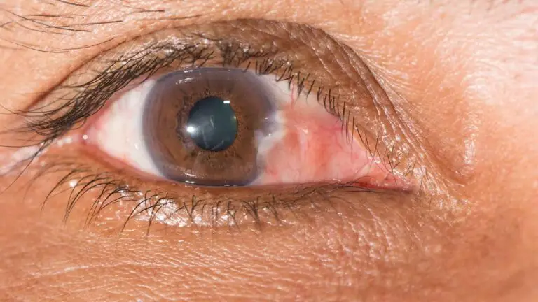
A pterygium crossing onto the cornea. After removal, the same area is where regrowth would begin, which is why it is covered with a graft.
How Recurrence Is Reduced
Covering the area with your own tissue.
Pterygium surgery with conjunctival autograft uses your own tissue to cover the area after removal. A thin graft of healthy conjunctiva, the membrane covering the white of the eye, is taken from under your upper eyelid and placed over the area.
A Cochrane review found this conjunctival autograft carried a lower risk of recurrence than amniotic membrane grafting, which uses donated tissue from the membrane surrounding a baby during pregnancy. The benefit was particularly clear in pterygia that had already recurred. ²
A report by the American Academy of Ophthalmology found that conjunctival autograft lowers recurrence compared with leaving the white of the eye bare. ³
Protection afterwards.
Light entering from the side of the eye may be focused onto the inner corner where pterygia form, ⁴ so wraparound sunglasses remain important after surgery.
Follow-up.
Your eye is reviewed after surgery so that any early regrowth is identified.

Repeat Surgery for Pterygium
Patients whose pterygium has returned after surgery elsewhere can usually still undergo repeat removal. Recurrent pterygia can be more firmly attached and scarred, so the assessment looks carefully at the cornea, the extent of the growth and the healthy conjunctiva available for grafting.
What to Expect
The operation is performed under local anaesthetic, which numbs the treatment area, and you go home the same day with drops.
- Return to work: most people return within three to seven days.
- Redness: this fades over two to six weeks.
- Settling: the eye fully settles over three to six months.
Many patients also have a pinguecula, a small, raised patch on the white of the eye. ⁵ This can be assessed at the same time.
Cost and Next Steps
Pterygium excision in Hatfield, with conjunctival autograft, is priced per eye on our costs page, and patient finance is available.
For further information, read the following pages:
Book a consultation with Blue Fin Vision® to discuss pterygium removal at our eye clinic at One Hatfield Hospital. Whether this is your first operation or the growth has returned, your consultant can assess your eye and explain your treatment options.
References
- Liu L, Wu J, Geng J, Yuan Z, Huang D. Geographical prevalence and risk factors for pterygium: a systematic review and meta-analysis. BMJ Open. 2013;3(11):e003787. doi:10.1136/bmjopen-2013-003787.
- Clearfield E, Muthappan V, Wang X, Kuo IC. Conjunctival autograft for pterygium. Cochrane Database Syst Rev. 2016;2(2):CD011349. doi:10.1002/14651858.CD011349.pub2.
- Kaufman SC, Jacobs DS, Lee WB, Deng SX, Rosenblatt MI, Shtein RM. Options and adjuvants in surgery for pterygium: a report by the American Academy of Ophthalmology. Ophthalmology. 2013;120(1):201-8.
- Coroneo MT. Pterygium as an early indicator of ultraviolet insolation: a hypothesis. Br J Ophthalmol. 1993;77(11):734-9.
- Panchapakesan J, Hourihan F, Mitchell P. Prevalence of pterygium and pinguecula: the Blue Mountains Eye Study. Aust N Z J Ophthalmol. 1998;26 Suppl 1:S2-5.
ABOUT THE AUTHOR
Mr Mfazo Hove
MBChB MD FRCOphth CertLRS
Consultant Ophthalmic Surgeon | Founder and Medical Director, Blue Fin Vision®
Mr Mfazo Hove is a Consultant Ophthalmic Surgeon with more than two decades of experience across NHS and private practice and more than 57,000 ophthalmic procedures performed. He trained for six and a half years at Moorfields Eye Hospital and served for five years as an NHS consultant at the Western Eye Hospital, Imperial College Healthcare NHS Trust.
He specialises in cataract surgery, refractive lens replacement, laser vision correction and implantable Collamer lenses (ICL). He is the founder and Medical Director of Blue Fin Vision®, a private ophthalmology group providing consultant-delivered care across London, Hertfordshire and Essex. His cataract surgery outcomes are publicly reported through the National Ophthalmology Database.
A ZEISS Key Opinion Leader and international speaker, Mr Hove is a contributing author to The Ophthalmologist and Ophthalmology Times. His writing examines how clinical governance and the organisation of ophthalmic services shape patients’ experiences and outcomes, with a focus on transparent outcome reporting, surgical training and long-term accountability for patient care.


