




Proliferative vs Non-Proliferative Diabetic Retinopathy: Understanding the Differences
- Posted
- Medically Reviewed by: Mr Mfazo Hove, Consultant Ophthalmic Surgeon
- Author: Mr Mfazo Hove
- Published: November 21, 2024
- Last Updated: November 10, 2026
TL;DR: In non-proliferative diabetic retinopathy (NPDR), blood vessels in the retina, the light-sensitive layer at the back of the eye, are damaged and leak, but no new vessels have formed. In proliferative diabetic retinopathy (PDR), parts of the retina have lost their blood supply, and fragile new vessels grow in response. Those new vessels can bleed or pull on the retina, so proliferative diabetic retinopathy needs treatment promptly, while non-proliferative diabetic retinopathy usually needs monitoring and good diabetes control.
The difference is whether the eye has started growing new vessels. Once it has, the priority changes from watching to treating.
Key Differences Between NPDR and PDR
Feature | Non-Proliferative Diabetic Retinopathy | Proliferative Diabetic Retinopathy |
|---|---|---|
What happens | Vessels leak and form microaneurysms, tiny bulges in weakened vessel walls, and small haemorrhages, areas of bleeding | New, fragile vessels grow on the retina or optic nerve, which carries visual signals to the brain |
Symptoms | Usually none, unless the macula, the central part of the retina responsible for detailed vision, swells | Often none at first; floaters, dots or lines in your vision, or sudden loss of vision if vessels bleed |
Main risks | Progression; macular oedema, swelling at the macula | Bleeding into the vitreous, the gel filling the eye; retinal detachment, where the retina pulls away from the back of the eye; a painful form of glaucoma, which can damage the optic nerve |
Typical management | Monitoring, diabetes and blood pressure control | Laser treatment, sometimes injections or surgery |
Both are defined on the international grading scale, and both arise from the same underlying damage to small blood vessels caused by diabetes.¹ ²
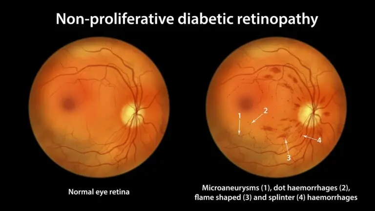
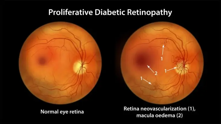
Why New Vessels Form
When enough capillaries, the smallest blood vessels, close, the starved retina releases growth signals, principally vascular endothelial growth factor (VEGF). New vessels grow in response, but they are fragile and grow in the wrong place, on the surface of the retina and into the vitreous gel.
Treating Proliferative Diabetic Retinopathy
- Laser, known as panretinal photocoagulation. Treating the peripheral retina, the area outside the centre, reduces the growth signal. The Diabetic Retinopathy Study showed it reduces the risk of severe vision loss by at least half in high-risk disease.³
- Anti-VEGF injections. These medicines block the growth signal and are used to control new vessels, alongside or instead of laser in selected patients.
- Vitrectomy. Surgery to clear blood that does not settle, or to release scar tissue pulling on the retina.
Slowing Progression
Intensive blood sugar control substantially reduces the progression of retinopathy.⁴
Blood pressure, cholesterol and stopping smoking matter too.² Macular oedema can occur at either stage, and our article on Diabetic Maculopathy and Retinopathy explains that distinction.
Warning Signs That Need Same-Day Attention
- A sudden shower of floaters, or a cobweb or red haze across your vision.
- Sudden blurring or loss of vision.
- A shadow spreading across part of your vision.
- A painful, red eye with blurred vision.
These can signal bleeding inside the eye, retinal detachment or raised eye pressure, and need assessment the same day.
Contact your urgent eye service immediately. If you cannot access it, ask for an urgent GP appointment or contact NHS 111. Do not wait for a routine consultation or your next screening appointment.⁵
If you suddenly cannot see from one or both eyes, or develop sudden severe eye pain, go to A&E or call 999. Do not drive yourself.⁵

Complications of Proliferative Retinopathy
Vitreous Haemorrhage
New vessels bleed into the vitreous gel. Vision can drop suddenly, from a few floaters to near-total blur. Small bleeds often clear over weeks; larger ones may need surgery.
Tractional Retinal Detachment
Scar tissue that forms around new vessels can contract and pull the retina away from the back of the eye. This needs vitreoretinal surgery, surgery involving the retina and the gel inside the eye.
Neovascular Glaucoma
New vessels can also grow on the iris, the coloured part of the eye, and block the eye’s drainage, causing a painful rise in pressure. It is serious, but less common when proliferative disease is treated promptly.
Can Non-Proliferative Diabetic Retinopathy Be Treated?
Most non-proliferative retinopathy needs no eye treatment. The priority is control of blood sugar, blood pressure and cholesterol, with monitoring so that progression, or swelling at the macula, is caught early.² ⁴ Severe non-proliferative disease is watched closely, and treatment is sometimes considered when the risk of progression is high.
What to Expect After Laser
Your vision may be blurred and dazzled for a few hours, so arrange a lift home. A mild ache is common and settles with simple pain relief. It can take several weeks for new vessels to regress, and some people need more than one session.⁴ ⁶
Vitrectomy, Explained
In a vitrectomy, the vitreous gel is removed, together with blood and scar tissue, and laser can be applied at the same time. It is performed in an operating theatre, usually under local anaesthetic, which numbs the eye while you remain awake, and is often a day case.
At Blue Fin Vision®, vitreoretinal surgery is performed by Professor Mahmut Dogramaci.
Frequently Asked Questions
Does non-proliferative always become proliferative?
No. Many people stay at non-proliferative stages for life, particularly with good control of diabetes.
Will laser cure proliferative retinopathy?
Laser controls new vessels and reduces the risk of severe sight loss by at least half in high-risk disease.³ It does not cure diabetes, so monitoring continues.
Are injections better than laser?
Both have a role. Injections act quickly and are used alongside laser in some patients; laser gives a longer-lasting effect without repeated injections. Your consultant will explain which suits your eye.
Can I exercise with proliferative retinopathy?
Most exercise is fine and good for your health. Ask your consultant about heavy straining or contact sport if you have active new vessels or recent bleeding.
At Blue Fin Vision®
At our eye clinic, we assess diabetic retinopathy with a full retinal examination and optical coherence tomography (OCT) imaging, a scan that produces detailed cross-sectional images of the retina.
Where injections are needed, they are given at our Harley Street clinic the same day or the following day, followed by an organised next step with us or through the NHS.
We plan for complications, with urgent assessment available at weekends. If you need vitrectomy, it is performed within the same team by Professor Mahmut Dogramaci, Consultant Vitreoretinal Surgeon.
These resources explain more about our service:
- Our article on Diabetic Maculopathy explains how the service works.
- Our Medical Retina page describes our care for conditions affecting the retina.
- Our Vitreoretinal Surgery page explains the surgical service.
Further patient information is available through:
- The Royal National Institute of Blind People (RNIB)’s free Understanding Eye Conditions Related to Diabetes guide.
- Our Patient Information.
Not sure what your diagnosis means for your sight? Book a consultation with Blue Fin Vision®. Our consultants will explain your retinal findings and whether you need monitoring or treatment, with clinics in London, Hertfordshire & Essex.
References
- Wilkinson CP, Ferris FL 3rd, Klein RE, Lee PP, Agardh CD, Davis M, et al. Proposed international clinical diabetic retinopathy and diabetic macular edema disease severity scales. Ophthalmology. 2003;110(9):1677-82.
- NHS. Diabetic retinopathy [Internet]. 2025 Jul 17 [cited 2026 Nov 8].
- Diabetic Retinopathy Study Research Group. Photocoagulation treatment of proliferative diabetic retinopathy. Clinical application of Diabetic Retinopathy Study (DRS) findings, DRS report number 8. Ophthalmology. 1981;88(7):583-600.
- National Institute for Health and Care Excellence. Diabetic retinopathy: management and monitoring. NICE guideline NG242. London: NICE; 2024.
- NHS. Vision loss [Internet]. 2025 Aug 28 [cited 2026 Nov 8].
- Kingston and Richmond NHS Foundation Trust. Panretinal photocoagulation (PRP) laser treatment [Internet]. 2025 Feb 17 [cited 2026 Nov 8].
ABOUT THE AUTHOR
Mr Mfazo Hove
MBChB MD FRCOphth CertLRS
Consultant Ophthalmic Surgeon | Founder and Medical Director, Blue Fin Vision®
Mr Mfazo Hove is a Consultant Ophthalmic Surgeon with more than two decades of experience across NHS and private practice and more than 57,000 ophthalmic procedures performed. He trained for six and a half years at Moorfields Eye Hospital and served for five years as an NHS consultant at the Western Eye Hospital, Imperial College Healthcare NHS Trust.
He specialises in cataract surgery, refractive lens replacement, laser vision correction and implantable Collamer lenses (ICL). He is the founder and Medical Director of Blue Fin Vision®, a private ophthalmology group providing consultant-delivered care across London, Hertfordshire and Essex. His cataract surgery outcomes are publicly reported through the National Ophthalmology Database.
A ZEISS Key Opinion Leader and international speaker, Mr Hove is a contributing author to The Ophthalmologist and Ophthalmology Times. His writing examines how clinical governance and the organisation of ophthalmic services shape patients’ experiences and outcomes, with a focus on transparent outcome reporting, surgical training and long-term accountability for patient care.