- Medically Reviewed by: Mr Mfazo Hove, Consultant Ophthalmic Surgeon
- Author: Mr Mfazo Hove
- Published: June 4, 2026
- Last Updated: June 12, 2026
Why It Is the Icing on the Cake, Not a Sign of Failure
There is a persistent misconception in cataract and refractive surgery: that needing an enhancement means the original operation has not worked. It is one of the most damaging narratives in the field, because it is wrong.
The reality is that premium cataract and refractive surgery is best understood as a two-stage optical process. Once that is clear, enhancement is no longer a complication or a rescue, it is refinement.
At Blue Fin Vision®, this distinction is critical. Not just clinically, but psychologically. How a patient interprets this phase often determines how they judge the entire outcome of their surgery.
Stage 1 - The Primary Operation: Precision, Not Perfection
The aim of the first stage is simple: get as close to the intended refractive target as possible. This is achieved through dual biometry, advanced diagnostics including OCT, corneal topography, and epithelial mapping where required, endothelial assessment, and modern IOL formula selection optimised against anatomy and visual goals.
Across the published literature, modern cataract surgery typically achieves around 70-80% of eyes within ±0.5 D and 90-95% within ±1.0 D of target¹ ². This range reflects inherent variability in axial length measurement, corneal power estimation, and effective lens position prediction² ³.
Across the Blue Fin Vision® 2024-2025 series, our outcomes sit above these benchmarks: approximately 98% of eyes are within ±1.0 D of target across all cases. Importantly, this reflects real-world practice. It is not adjusted to exclude complex eyes, including previous laser eye surgery, radial keratotomy, keratoconus, highly myopic eyes, or atypical corneal anatomy.
It reflects a system built on measurement redundancy, advanced diagnostics, meticulous planning, and fully consultant-delivered surgery.
When Predictability Falls: Eyes That Behave Differently
A significant proportion of patients we treat have been declined elsewhere. They are not just technically harder; they are less predictable by definition.
Keratoconus and Irregular Corneas
Irregular corneal optics reduce the accuracy of intraocular lens power calculations even with modern keratoconus-adjusted formulae⁷.
Post-RK Corneas
Corneal biomechanics are altered, and refractive outcomes remain inherently variable following cataract surgery⁵.
Highly Myopic Eyes
Axial length extremes reduce refractive predictability despite optimisation strategies⁴.
Flat Corneas
When keratometry deviates significantly from normal ranges, effective lens position assumptions become less reliable³.

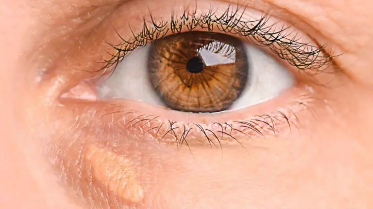
Figure 1. Bilateral flat corneas with K1 values of 32.12 D and 32.06 D, well below the normal range of approximately 41-46 D. The kind of biometry routinely declined elsewhere, and the kind we routinely treat.
Precision remains high, but predictability falls.
Stage 2 - Enhancement: Defined, Not Reactive
At Blue Fin Vision®, enhancement is not improvised. It is defined by a clear clinical threshold:
Blue Fin Vision® Enhancement Criterion
Enhancement is offered when residual refractive error is ≥1.0 dioptre spherical equivalent from the intended target, subject to clinical suitability.
Because Stage 1 accuracy is high, approximately 98% of patients fall within this threshold and around 2% require refinement. This 2% rate includes all cases; we do not exclude complex eyes from our data. Even with optimal planning, residual refractive error remains an expected part of cataract and refractive surgery due to biological variability² ⁸.
Establishing the Enhancement Pathway at the First Consultation
The first consultation is not only for planning Stage 1. It also determines what Stage 2 will look like, if it is required.
Every patient at Blue Fin Vision® is assessed for laser eye surgery suitability at the first consultation as standard. This forms part of the standard diagnostic workup, alongside biometry, corneal topography, and endothelial assessment, and it defines which enhancement pathway is available. The patient is advised accordingly.
If laser eye surgery is viable, corneal enhancement is the most accurate method of refining low-grade residual refractive error. If laser is not viable, thin corneas, altered post-RK biomechanics, irregular topography, established keratoconus, or other contraindications, the enhancement plan must be lens-based from the outset, typically through a piggyback intraocular lens or a staged refractive strategy⁶ ¹⁰. In every case, the patient is told what is on the table for them, what the alternative is, and why. The two-stage plan is therefore designed at the first consultation, not deferred until after surgery.
The system does not rely on Stage 2 being universally or reliably available. It is built around knowing, in advance, what Stage 2 will be. We call this Stage Zero: the moment the enhancement pathway is decided, before the first procedure is performed.
This is what allows the Blue Fin Vision® system to maintain its accuracy across both routine and complex eyes.
Stage 2 is decided at Stage Zero.
Why Enhancement Is Part of the Plan
In routine eyes, enhancement is refinement. In complex eyes, it is often anticipated from the outset, through piggyback intraocular lenses for residual refractive error, staged refractive strategies in post-RK patients, situations where lens platform limitations prevent full correction in a single step, and irregular corneas including keratoconus.
Piggyback intraocular lenses are a recognised and established method for correcting residual refractive error when corneal laser enhancement is not suitable⁶ ¹⁰. In these patients, the surgery is not incomplete without enhancement, the plan is incomplete without it. These cases are planned and consented as two-stage procedures from the outset, with all surgery performed by the named consultant, Mr Mfazo Hove.
The Conversation Most Providers Avoid: Cost in Complex Cases
Complex eyes require longer consultations, often more than one, additional diagnostics, intraoperative flexibility, backup strategies, and greater surgical experience. Sometimes they require staged procedures, multiple admissions, or piggyback intraocular lenses.
Charging more for complex cases reflects the additional time and expertise required, but creates a dangerous assumption: that paying more guarantees a perfect outcome. Biology does not work that way.
Standardise pricing.
Absorb complexity.
This is the Blue Fin Vision® approach.
The focus remains on delivering the best possible outcome for every patient, regardless of complexity. Patients are not penalised for previous surgery, irregular corneas, or complex anatomy. The system absorbs the additional workload, planning is extended where required, and access to refinement is maintained.
The most complex cases are often the least profitable, but they are the ones that matter most.
The Psychological Gap
Patients are often told: “We aim to get you glasses-free.” What they hear is: “One surgery will achieve everything.” So, when enhancement is mentioned, it feels like failure. When in reality, it is completion.
What Actually Defines Quality
Not perfection, but preparation. At Blue Fin Vision®, quality is defined by depth of consultation, redundancy in measurement, structured planning, access to enhancement pathways, and clear communication throughout.
The true measure of a system is not how it performs when everything goes right, but how it is designed for when outcomes are less predictable² ⁹.
See what our patients say. Read more here.
Final Thought
Enhancement is not a sign that surgery has failed. It is the final step in delivering the best possible optical outcome. And in complex eyes, keratoconus, post-RK, extreme myopia, it is often part of the journey from the beginning.
The reward is not in simplicity.
It is in having a system capable of managing complexity and finishing the job properly.
Outcome data referenced in this piece is published in full on the Blue Fin Vision® Advantage page.
References
- Hoffer KJ. The Hoffer Q formula: a comparison of theoretic and regression formulas. Journal of Cataract and Refractive Surgery. 1993;19(6):700-712.
- Norrby S. Sources of error in intraocular lens power calculation. Journal of Cataract and Refractive Surgery. 2008;34(3):368-376.
- Olsen T. Calculation of intraocular lens power: a review. Acta Ophthalmologica Scandinavica. 2007;85(5):472-485.
- Wang L, Koch DD. Modified axial length adjustment formulas in long eyes. Journal of Cataract and Refractive Surgery. 2018;44(11):1396-1397.
- Turnbull AMJ, Crawford GJ, Barrett GD. Methods for intraocular lens power calculation in cataract surgery after radial keratotomy. Ophthalmology. 2020;127(1):45-51.
- Gayton JL, Sanders V, Van der Karr M, Raanan MG. Piggybacking intraocular implants to correct pseudophakic refractive error. Ophthalmology. 1999;106(1):56-59.
- Kane JX, Connell B, Yip H, McAlister JC, Beckingsale P, Snibson GR, Chan E. Accuracy of intraocular lens power formulas modified for patients with keratoconus. Ophthalmology. 2020;127(8):1037-1042.
- Aristodemou P, Knox Cartwright NE, Sparrow JM, Johnston RL. Intraocular lens formula constant optimization and partial coherence interferometry biometry: refractive outcomes in 8108 eyes after cataract surgery. Journal of Cataract and Refractive Surgery. 2011;37(1):50-62.
- Cooke DL, Cooke TL. Comparison of 9 intraocular lens power calculation formulas. Journal of Cataract and Refractive Surgery. 2016;42(8):1157-1164.
- Sáles CS, Manche EE. Managing residual refractive error after cataract surgery. Journal of Cataract and Refractive Surgery. 2015;41(6):1289-1299.
ABOUT THE AUTHOR
Mr Mfazo Hove
Consultant Ophthalmic Surgeon
MBChB MD FRCOphth CertLRS
Mr Mfazo Hove is a Consultant Ophthalmic Surgeon with experience spanning more than 57,000 procedures. He completed 6.5 years of specialist training at Moorfields Eye Hospital and served for five years as a consultant at the Western Eye Hospital, Imperial College Healthcare NHS Trust. He is the founder of Blue Fin Vision®, a consultant-led private ophthalmology practice operating across London, Essex, and Hertfordshire. His clinical expertise encompasses advanced cataract surgery, refractive lens replacement, laser vision correction, and implantable Collamer lenses (ICL).
A ZEISS Key Opinion Leader, Mr Hove is a respected international speaker with five invited engagements across seven cities in 2026:
- ZEISS China tour (Changsha, Shanghai, and Hangzhou, April – ZEISS APAC User Meeting)
- RCOphth Annual Congress – May – Manchester
- ZEISS EMEA User Meeting (Istanbul)
- ZEISS Lausanne User Meeting (Lausanne)
- European Society of Cataract and Refractive Surgeons Annual Congress (ESCRS, London)
Related Topics
Understanding Your Refractive Outcome
Setting Realistic Expectations
The Consultation and Patient Satisfaction
Schedule Your Consultation Today
If you have been told your eyes are too complex to treat, or you simply want clarity on what a two-stage plan would mean for you, the Blue Fin Vision® team is here to help. You would be trusting your vision to a consultant-led UK clinic with documented outcomes and a network of centres across London, Hertfordshire, and Essex, offering greater access to Harley Street standards. Book a consultation or discuss your options with our team today.