



Patient Experience
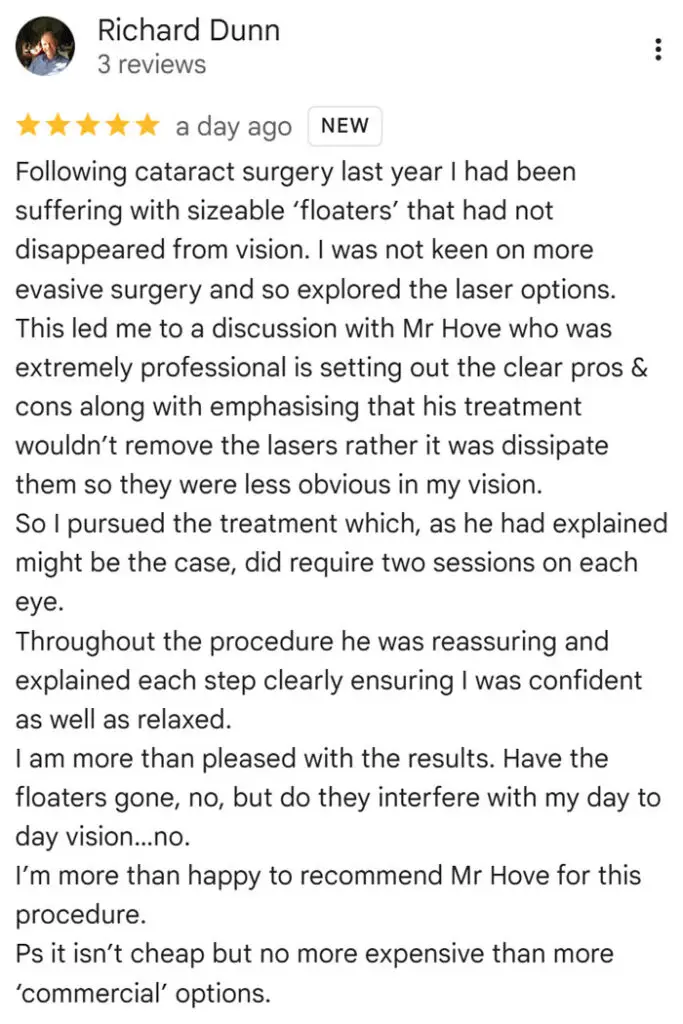
“Following cataract surgery last year I had been suffering with sizeable ‘floaters’ that had not disappeared from vision. I was not keen on more evasive surgery and so explored the laser options.
This led me to a discussion with Mr Hove who was extremely professional is setting out the clear pros & cons along with emphasising that his treatment wouldn’t remove the lasers rather it was dissipate them so they were less obvious in my vision.
So I pursued the treatment which, as he had explained might be the case, did require two sessions on each eye.
Throughout the procedure he was reassuring and explained each step clearly ensuring I was confident as well as relaxed.
I am more than pleased with the results. Have the floaters gone, no, but do they interfere with my day to day vision…no.
I’m more than happy to recommend Mr Hove for this procedure.
Ps it isn’t cheap but no more expensive than more ‘commercial’ options.”
Clinical Explanation
New floaters after cataract surgery are most often caused by a posterior vitreous detachment (PVD). The vitreous is the clear gel that fills the back of the eye. With age it becomes more liquid and eventually peels away from the retina, the light-sensitive layer at the back of the eye.
As it separates, condensed strands of gel, and the ring of gel that was attached around the optic nerve, which carries visual signals to the brain, cast shadows on the retina. These shadows are seen as floaters: dots, cobwebs, rings or larger drifting shapes.
Some people also notice brief flashes of light as the gel tugs on the retina.
Cataract surgery brings this process forward. In a study of patients aged 50 to 60, a PVD was found in 50.8% of eyes that had undergone cataract surgery, compared with 20.8% of the fellow eyes that had not, and the rate was higher in longer, more short-sighted eyes.¹
New floaters in the months or years after surgery are therefore common. In most cases they are the vitreous doing what it would have done anyway, only sooner.
Not every floater noticed after surgery is new. A cataract scatters and dims the light entering the eye, so floaters that were already there can become far more obvious once vision is clear again. Whether a floater is new or newly noticed cannot be told from the symptom alone, which is why both are examined rather than assumed to be harmless.
The gel does not always separate cleanly. Where it is firmly attached, it can pull a tear in the retina as it comes away. A retinal tear can allow fluid to pass under the retina and lift it off, a retinal detachment, which threatens sight. This is why new floaters are never simply reassured over the phone.
Structured Context
This applies to you if: you have had cataract or lens replacement surgery and notice new floaters, a sudden increase in floaters, flashes of light, a shadow or curtain moving across your vision, or new blurring.
This does not apply to:
- Floaters you have had unchanged for years, from before your surgery and already examined.
- Floaters linked to inflammation inside the eye or to bleeding from conditions such as diabetic retinopathy, damage to the retina’s blood vessels caused by diabetes, which need a different assessment.
How quickly to be seen: a sudden shower of floaters, flashes, a curtain or shadow, or a drop in vision needs urgent examination, ideally the same day.
Blue Fin Vision® has seven-day emergency retinal capability under Professor Mahmut Dogramaci, Consultant Vitreoretinal Surgeon, a specialist in conditions affecting the retina and vitreous gel.
What the check involves: the pupil is dilated, meaning widened with eye drops, and the whole retina is examined, including the far periphery, its outermost edges, where most tears occur.
The gel behind the lens is also examined for pigment or blood, which raise the likelihood of a tear. Because a lens implant gives a clear view, this examination is usually straightforward after cataract surgery.
If the retina is healthy: the floaters are left alone in the first instance.
A normal first examination does not rule out a later tear, however: in one review, 3.4% of patients whose first examination showed an uncomplicated PVD, meaning no associated retinal tear or other complication was found, developed a tear within six weeks.²
Return for urgent assessment, the same day, if your floaters increase, flashes start or worsen, your vision becomes blurred, or a shadow or curtain appears.
You will be told at your examination whether a repeat check is needed and when.
If the floaters persist: if they continue to interfere with daily life once the retina has been confirmed healthy, the options are explained in detail on our page on YAG laser vitreolysis for floaters after cataract surgery. YAG laser vitreolysis is a laser treatment intended to make floaters less noticeable.
Published Evidence
Cataract surgery makes PVD more likely. In a comparative series of 149 patients aged 50 to 60, PVD was documented in 50.8% of operated eyes and 20.8% of unoperated fellow eyes, with higher rates in eyes 25 mm or longer.¹
New floaters carry a real risk of retinal tear. A systematic review of 17 studies in JAMA found that among patients with sudden-onset floaters or flashes seen by an ophthalmologist, an eye specialist, 14% had a retinal tear. Reduced vision was the symptom most strongly linked to a tear.²
Patients whose first examination showed an uncomplicated PVD still had a 3.4% chance of a tear appearing within six weeks, with the risk rising if 10 or more new floaters or reduced vision developed.²
What the examiner sees matters. Patients with pigment or blood in the vitreous were 52 times more likely to have a retinal tear than those whose vitreous looked normal.³
The rates in routine care. In 8,305 patients with acute PVD, meaning a recent-onset PVD, seen in a large comprehensive eye care system, 5.4% had a retinal tear and 4% a retinal detachment at their first examination.⁴
Retinal detachment after cataract surgery is uncommon but real. In a whole-population study of 65,055 cataract operations, the 10-year cumulative incidence of retinal detachment, meaning the estimated proportion developing it over ten years, was 0.68%, with higher risk in patients under 60 and in men.⁵
Treatment options for persistent floaters. The two interventions used for floaters that do not settle are YAG laser vitreolysis and vitrectomy, surgery to remove the vitreous gel. A Cochrane review found no randomised trials comparing the two directly.⁶
Surgeon Interpretation
New floaters after cataract surgery should never be ignored. In most patients they come from a posterior vitreous detachment, which is common and usually harmless, but a small number have a retinal tear, and a tear found early can be treated before it becomes a detachment. So the first step is always a dilated retinal examination, not reassurance.
Once the retina is confirmed healthy, I tell patients that most people learn to ignore the new floaters. They become less noticeable over the following weeks and months, and for most patients that is the end of it.
For the minority whose floaters remain intrusive, there are three options: leave them alone, YAG laser vitreolysis, or vitrectomy.
Vitrectomy is available on the NHS and privately. YAG vitreolysis is a private-only option.
Laser vitreolysis breaks large floaters into smaller ones that are easier to ignore, but it does not remove them.
Richard’s floaters were large and had persisted for months, which is the point at which that conversation becomes worthwhile.
Mr Mfazo Hove, Consultant Ophthalmic Surgeon.
Clinical Takeaway
New floaters after cataract surgery are usually a posterior vitreous detachment, but every new floater needs a dilated retinal examination before anything else, because a small proportion signal a retinal tear.
References
- Hilford D, Hilford M, Mathew A, Polkinghorne PJ. Posterior vitreous detachment following cataract surgery. Eye (Lond). 2009;23(6):1388-92.
- Hollands H, Johnson D, Brox AC, Almeida D, Simel DL, Sharma S. Acute-onset floaters and flashes: is this patient at risk for retinal detachment? JAMA. 2009;302(20):2243-9.
- Sharma S, Walker R, Brown GC, Cruess AF. The importance of qualitative vitreous examination in patients with acute posterior vitreous detachment. Arch Ophthalmol. 1999;117(3):343-6.
- Seider MI, Conell C, Melles RB. Complications of acute posterior vitreous detachment. Ophthalmology. 2022;129(1):67-72.
- Clark A, Morlet N, Ng JQ, Preen DB, Semmens JB. Risk for retinal detachment after phacoemulsification: a whole-population study of cataract surgery outcomes. Arch Ophthalmol. 2012;130(7):882-8.
- Kokavec J, Wu Z, Sherwin JC, Ang AJS, Ang GS. Nd:YAG laser vitreolysis versus pars plana vitrectomy for vitreous floaters. Cochrane Database Syst Rev. 2017;6(6):CD011676.


