Patient Experience
“After several months of suffering with severe redness, swelling, and scaling of the eyelids, I found Dr. Mfazo Hove on 11 April 2026. He immediately diagnosed me with allergic conjunctivitis and eczematous eyelids. Dr. Hove is an incredibly kind and compassionate specialist who truly understood how much this condition was affecting my quality of life. He reassured me that I would see a visible improvement within two weeks and a full resolution within a month and he was absolutely right. Three weeks into my treatment, my eyes look and feel so much better, and I finally feel like my old self again. Dr. Hove sees you as a person, not just a patient, and his follow-up care is both professional and deeply humane. I cannot recommend him highly enough for his exceptional care.”
Chronic eyelid eczema with allergic conjunctivitis can be fully resolved within four weeks, when both compartments are treated correctly.
Clinical Images: Before Treatment and at Three Weeks
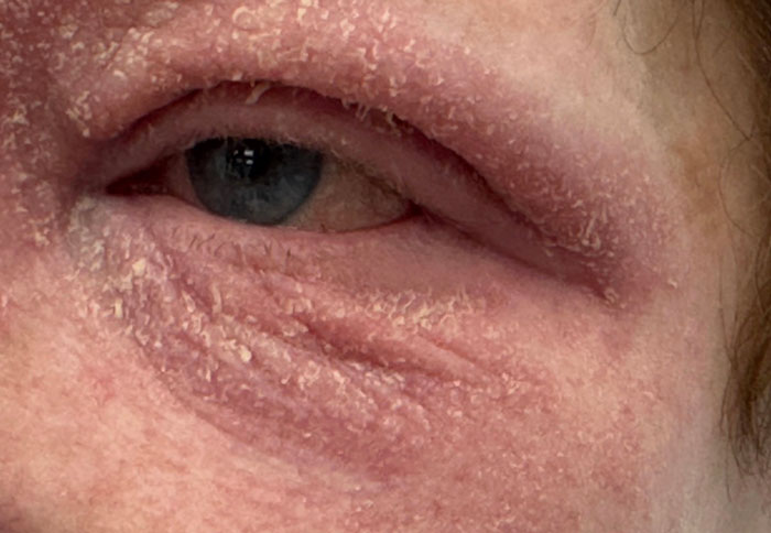
Figure 1. Severe periocular eczema at presentation: dense scaling, fissuring, and erythema involving the upper and lower eyelid skin and adjacent periorbital region.
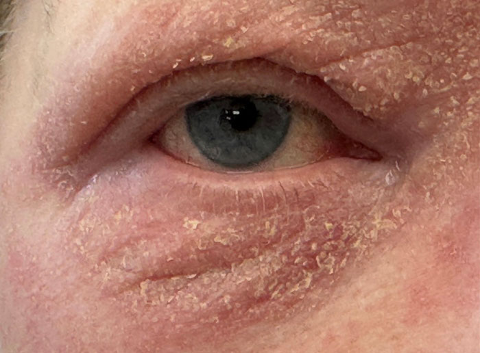
Figure 2. Severe periocular eczema at presentation, second view: confirms the full extent of eyelid skin involvement, with associated conjunctival inflammation visible on the ocular surface.
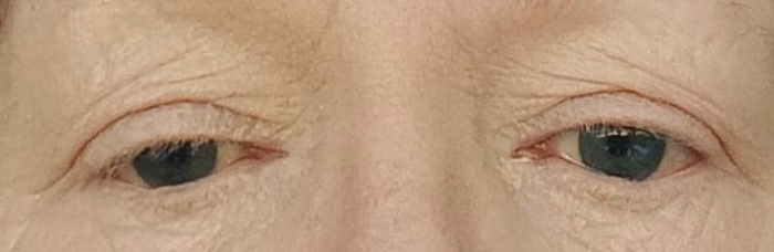
Figure 3. Near-complete resolution of eyelid skin and conjunctival surface at three weeks of dual-compartment topical therapy.
The visible change between Figures 1 to 2 and Figure 3 represents the expected response when both inflammatory compartments are treated simultaneously.
What Was Actually Happening
As shown in Figures 1 and 2, the presenting picture combined two coexisting conditions that frequently occur together but are routinely treated as one. The conjunctival surface showed allergic conjunctivitis, redness, lid oedema, watering, ocular irritation, driven by an IgE-mediated hypersensitivity response on the conjunctival mucosa. The eyelid skin showed eczematous dermatitis, fine scaling, fissuring, erythema, chronic itch, reflecting a barrier-disrupted, T-cell-mediated inflammation of the periocular skin.
This is not a single disease. It is a linked inflammatory system across two anatomical compartments, the conjunctival mucosa (IgE-mediated hypersensitivity) and the eyelid skin (barrier-disrupted, T-cell-mediated dermatitis). Treating one while leaving the other active guarantees recurrence.
Correct treatment therefore requires both surfaces to be quietened simultaneously, a dual-action allergy eye drop on the ocular surface, alongside a topical corticosteroid skin preparation prescribed at a potency appropriate for eyelid skin and used for a defined, supervised course. This is the principle behind the resolution shown in Figure 3.
The diagnosis is clinical and can be made at the first consultation without laboratory testing in typical cases.
Why This Condition Often Persists for Months
Most patients have already been treated before they arrive. The pattern is consistent:
- Allergy eye drops alone, temporary relief, rapid recurrence.
- Topical skin treatment alone, partial improvement, persistent irritation.
- No defined timeline, chronic cycling disease.
The underlying issue is not severity. It is incomplete treatment of a dual-compartment disease. Patients in this position do not need stronger therapy, they need both compartments addressed at once, on a defined timeline, with scheduled review.
Who This Proof Page Applies To
This pattern of resolution applies to:
- Adults with chronic redness, swelling, and scaling of the eyelid skin coexisting with conjunctival inflammation.
- Patients with a personal or family history of atopy, hay fever, eczema, or asthma.
- Patients in whom previous single-agent therapy (eye drops alone, or skin cream alone) has failed to give durable resolution.
- Adults whose disease is functional, affecting reading, screen tolerance, sleep, or quality of life, rather than purely cosmetic.
Who This Does Not Apply To
- Patients with infectious blepharitis (bacterial, viral, or demodex), where antimicrobial or lid-hygiene management takes priority over an anti-inflammatory pathway.
- Patients with contact dermatitis from a specific cosmetic, eye drop preservative, nail product, or hair dye, these require trigger identification and removal rather than chronic topical steroid.
- Patients with eyelid skin lesions of suspicious morphology, asymmetry, or recent change, these should be assessed for alternative diagnoses before periocular steroid therapy is initiated.
- Patients with significant corneal involvement (atopic keratoconjunctivitis with corneal staining or shield ulceration), these require closer surveillance and a more nuanced ladder of therapy.
Expected Treatment Timeline
When the diagnosis is correct on day one and both compartments are treated in parallel on a defined course, the following trajectory is the expected outcome rather than an exceptional one:
- Week 1 to 2: Reduction in redness, itch, and scaling. Conjunctival reaction begins to settle.
- Week 2 to 3: Visible improvement in eyelid skin integrity. Patient reports return of comfort and screen tolerance.
- Week 4: Full resolution in typical cases. Topical corticosteroid is stopped on a defined taper; ocular surface treatment reviewed.
What the Published Evidence Says
Allergic conjunctivitis is the most common immunological disease of the ocular surface, affecting an estimated 15 to 20% of the general population, and is formally classified as a hypersensitivity disorder of the conjunctiva by the International Ocular Inflammation Society and EAACI.¹ The diagnostic features, bilateral redness, lid oedema, itch, and conjunctival reaction, are recognisable on slit-lamp examination without the need for laboratory testing in the typical case.
Eczematous involvement of the eyelid skin is a recognised periocular manifestation of atopic disease and overlaps mechanistically with atopic dermatitis elsewhere, with shared T-helper-2 cytokine signalling and skin barrier dysfunction.² ³ The eyelid is the thinnest skin on the body, which makes it both the first to flare and the most responsive to correctly chosen topical therapy.
Topical corticosteroids remain the cornerstone of acute control for eczematous eyelid disease in international consensus guidelines, used at appropriate potency for eyelid skin and for a defined course rather than open-ended use.⁴ The European Task Force on Atopic Dermatitis specifies that periocular skin tolerates short courses of low-to-mid-potency corticosteroid well when prescribed and supervised, and that under-treatment, not over-treatment, is the more common cause of chronic disease at this site.
Second-generation topical antihistamines and dual-action mast-cell stabiliser preparations have demonstrated rapid symptom relief in randomised trials for allergic conjunctivitis, with a clinical improvement signal typically established within 7 to 14 days of consistent use.⁵ This is consistent with the two-week visible-improvement marker forecast at the initial consultation and confirmed at follow-up.
Surgeon Interpretation
The clinically important point in this case is not that the patient improved. It is that the improvement was forecast at the first consultation and matched the published evidence to the week. That predictability is only possible when two things hold: the diagnosis is correct on day one, and both compartments, conjunctival surface and eyelid skin, are addressed at the same time.
Patients with this presentation often arrive having been treated for one half of the disease in isolation. They have been given allergy drops without skin treatment, or a skin cream without an eye drop, and the residual untreated compartment continues to drive recurrence. The resulting pattern, months of low-grade flare with intermittent partial relief, is what this patient described in the opening line of her review.
The treatment principle is straightforward but precise. A dual-action allergy eye drop quietens the conjunctival mast cell and histamine response. A periocular topical corticosteroid, prescribed at a potency appropriate for eyelid skin and used for a short defined course with a clear stop point, restores barrier function and breaks the inflammatory loop. Both are reviewed at follow-up rather than left open-ended.
This is the principle behind the Blue Fin Vision® approach to medical ophthalmology: accurate diagnosis at the first consultation, defined treatment pathways, and scheduled review rather than open-ended therapy.
When that framework is applied, resolution is predictable, not exceptional.
Clinical Takeaway
Severe eyelid eczema with allergic conjunctivitis is a dual-compartment disease. Treated as one, a dual-action allergy eye drop on the ocular surface and a periocular topical corticosteroid on the eyelid skin, supervised on a defined timeline, visible improvement is expected within two weeks and full resolution within a month. Months of partial-relief treatment is not a failure of the patient. It is a failure to treat both compartments simultaneously.
References
- Leonardi A, Bogacka E, Fauquert JL, Kowalski ML, Groblewska A, Jedrzejczak-Czechowicz M, Doan S, Marmouz F, Demoly P, Delgado L. Ocular allergy: recognizing and diagnosing hypersensitivity disorders of the ocular surface. Allergy. 2012;67(11):1327-1337.
- Bielory L. Allergic and immunologic disorders of the eye. Part II: ocular allergy. Journal of Allergy and Clinical Immunology. 2000;106(6):1019-1032.
- Foster CS, Calonge M. Atopic keratoconjunctivitis. Ophthalmology. 1990;97(8):992-1000.
- Wollenberg A, Barbarot S, Bieber T, Christen-Zaech S, Deleuran M, Fink-Wagner A, Gieler U, Girolomoni G, Lau S, Muraro A, Czarnecka-Operacz M, Schäfer T, Schmid-Grendelmeier P, Simon D, Szalai Z, Szepietowski JC, Taïeb A, Torrelo A, Werfel T, Ring J. Consensus-based European guidelines for treatment of atopic eczema (atopic dermatitis) in adults and children: part I. Journal of the European Academy of Dermatology and Venereology. 2018;32(5):657-682.
- Bielory L, Lien KW, Bigelsen S. Efficacy and tolerability of newer antihistamines in the treatment of allergic conjunctivitis. Drugs. 2005;65(2):215-228.


