- Medically Reviewed by: Mr Mfazo Hove, Consultant Ophthalmic Surgeon
- Author: Mr Mfazo Hove
- Published: February 16, 2026
- Last Updated: May 1, 2026
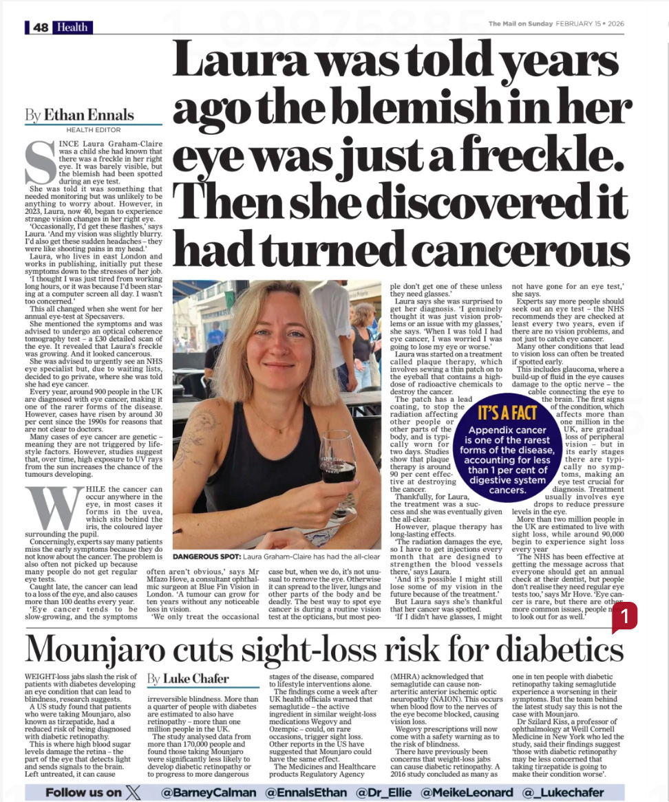
Most people understand what a freckle on the skin represents. But when a patient is told they have a “freckle in the eye”, the psychological impact is very different, largely because a minority of pigmented ocular lesions are associated with melanoma.
The important message is reassuring: most ocular “freckles” are benign. The equally important message is clinical: they must be correctly classified and properly monitored.
The difference between harmless pigment and early malignancy is not intuition, it is structured examination supported by imaging and validated risk models.
What does "freckle in the eye" actually mean?
Patients use this term to describe several distinct entities.
- Choroidal naevus
A pigmented lesion beneath the retina in the choroid. Often detected incidentally during dilated retinal examination. Most remain stable. A small proportion transform into choroidal melanoma over time. ¹
- Iris naevus
Pigment on the coloured iris surface. Usually benign but long-term growth into iris melanoma has been documented when specific risk features are present. ²
- Conjunctival naevus
Pigment on the surface membrane covering the white of the eye. Common in childhood or early adulthood and often shows intralesional cysts. Malignant transformation is uncommon. ³
- Primary Acquired Melanosis (PAM)
Flat patchy surface pigmentation typically appearing in adulthood. Malignant potential depends heavily on histological atypia and lesion extent. ⁴
The term “freckle” describes appearance, not behaviour. Clinical behaviour determines significance.
For a broader overview of what an eye freckle is and when treatment applies, read What is an Eye Freckle.
When should you worry?
The principle is simple:
Concerning lesions demonstrate objective signs of activity.
These include:
- Documented growth
- Thickness greater than expected
- Subretinal fluid
- Orange pigment (lipofuscin)
- New intrinsic or feeder vessels
- Tissue distortion
- Angle involvement (iris lesions)
- Nodularity or thickening (surface pigment)
These risk features have been validated in large cohorts and predict biological activity rather than cosmetic pigment. ¹ ²

Choroidal Naevus: The TFSOM-DIM Model
For choroidal lesions, the most validated transformation predictors are summarised by the TFSOM model:
- Thickness >2 mm
- Fluid (subretinal)
- Symptoms
- Orange pigment
- Margin near disc
Additional imaging features such as ultrasound hollowness and absence of halo or drusen further refine risk. ¹
In Shields’ landmark series of 2,514 cases, transformation risk rose sharply as these features accumulated. ¹
A flat lesion without risk factors has low short-term transformation probability. A lesion with multiple risk markers demands escalation.
Iris Lesions: ABCDEF Risk Guide
Iris naevi are common. Most are stable. However, long-term data show growth risk increases when the following are present:
- Age ≤40
- Blood (hyphema)
- Clock-hour inferior
- Diffuse
- Ectropion uveae
- Feathery margins
These parameters were derived from 1,611 consecutive eyes. ²
Pupil distortion, elevated pressure, intrinsic vessels, or documented enlargement are additional red flags.
Conjunctival Pigment: Location Matters
Surface pigment is usually benign, especially cystic interpalpebral conjunctival naevi. ³
Concern rises with:
- Atypical locations (fornix, caruncle, tarsal conjunctiva)
- Lack of cysts
- Rapid change
- Feeder vessels
- Nodularity
- Patchy spreading pattern (suggestive of PAM)
In PAM, risk correlates with histologic atypia and lesion clock-hour extent. ⁴
Imaging: Why Structure Matters
Assessment is multimodal:
- Colour photography (baseline comparison)
- OCT (detect fluid)
- Autofluorescence (orange pigment)
- B-scan ultrasound (thickness)
Modern practice relies on structured documentation rather than verbal reassurance. ⁵ ⁶
Learn more about how OCT imaging supports retinal assessment in What is a Posterior Segment OCT Eye Test?

What Symptoms Increase Urgency?
Seek reassessment if you develop:
- New central blur
- Distortion
- Scotoma
- Hyphema
- Sudden thickening of surface lesion
- Pain with iris distortion
Symptoms alone do not confirm malignancy, but they alter urgency. ⁷
Monitoring: Not Casual, But Protocol
Proper monitoring includes:
- Baseline imaging
- Objective measurement
- Risk scoring
- Defined recall interval
- Clear written documentation
Early detection of small melanomas improves outcomes. ⁷ ⁸
Most lesions remain stable. The system is built to identify the minority that evolve.
FAQ
Is a freckle in the eye usually cancer?
No. The vast majority are benign. Surveillance exists to protect patients from the minority that transform.
For a detailed explanation of ocular malignancies, read What is Eye Cancer? Understanding Ocular Malignancies.
Can I see it in the mirror?
Choroidal lesions cannot be seen without dilation. Surface lesions may be visible but require examination for risk features.
Should I remove it for peace of mind?
Removal is reserved for suspicion, growth, symptoms, atypical location, or diagnostic uncertainty. Routine excision is not recommended. ³
How often is follow-up needed?
It depends on risk markers. Low-risk lesions are reviewed periodically; higher-risk lesions require shorter intervals. ¹
What is the difference between a naevus and melanoma?
Melanomas demonstrate biological activity, thickness, fluid, orange pigment, vascular change, or documented progressive enlargement. ¹ ²
As Featured in the Daily Mail, Eye Cancer Commentary (February 2026)
Commentary by Mr Mfazo Hove, Consultant Ophthalmologist and Founder of Blue Fin Vision®
Read verified patient reviews on the Wall of Love.
References
- Shields CL, Furuta M, Berman EL, Zahler JD, Hoberman DM, Dinh DH, et al. Choroidal nevus transformation into melanoma: analysis of 2514 consecutive cases. Arch Ophthalmol. 2009;127(8):981-987. PMID: 19667334.
- Shields CL, Kaliki S, Hutchinson A, Nickerson S, Patel J, Kancherla S, et al. Iris nevus growth into melanoma: analysis of 1611 consecutive eyes. Ophthalmology. 2013;120(4):766-772. PMID: 23290981.
- Huang JJ, Locatelli EVT, Chocron A, Camacho MR, Dubovy S, Karp CL, et al. Conjunctival nevus. Curr Ophthalmol Rep. 2023;11(4):104-112. PMID: 38390435.
- Shields JA, Shields CL, Mashayekhi A, Marr BP, Benavides R, Thangappan A, et al. Primary acquired melanosis of the conjunctiva. Trans Am Ophthalmol Soc. 2007;105:61-71. PMID: 18427595.
- Dalvin LA, Shields CL, Ancona-Lezama D, Yu MD, et al. Multimodal imaging features predictive of choroidal nevus transformation. Br J Ophthalmol. 2019;103(10):1441-1447. PMID: 30523045.
- Pavlin CJ, Harasiewicz K, Sherar MD, Foster FS. Clinical use of ultrasound biomicroscopy. Ophthalmology. 1991;98(3):287-295. PMID: 2023747.
- Shields CL, Demirci H, Materin MA, Marr BP, Mashayekhi A, Shields JA. Clinical factors in identification of small choroidal melanoma. Can J Ophthalmol. 2004;39(4):351-357. PMID: 15327099.
- Shields CL, Pirondini C, Bianciotto C, et al. Autofluorescence of choroidal nevus. Retina. 2008;28(8):1035-1043. PMID: 18779708.
- Dalvin LA, Salomão DR, Patel SV. Incidence of conjunctival tumours. Br J Ophthalmol. 2018;102(12):1728-1734. PMID: 29511061.
- Chien JL, Sioufi K, Surakiatchanukul T, Shields CL. Choroidal nevus: review. Curr Opin Ophthalmol. 2017;28(3):228-237. PMID: 28141766.
Related Topics
Types of Eye Freckle
- What Does a “Freckle in the Eye” Actually Mean? Choroidal, Iris and Conjunctival Naevi Explained
- Iris “Freckles”: Using the ABCDEF Guide to Spot Higher-Risk Iris Naevi
- Conjunctival “Freckles” on the White of the Eye: When a Naevus Is Usually Harmless
- Primary Acquired Melanosis (PAM): Why Some Flat Conjunctival Pigment Needs Oncology Input
Risk Features and Scoring
- Choroidal Naevus vs Choroidal Melanoma: Understanding TFSOM-DIM Risk Factors
- MOLES and TFSOM-DIM: How Specialists Score Choroidal Naevi and Decide on Referral
- Growth Without Transformation: When a Choroidal Naevus Gets Bigger but Isn’t Melanoma
- High-Risk Features in Conjunctival Pigmented Lesions: PAM, Forniceal Location and Feeder Vessels
Symptoms and Urgency
Imaging and Assessment
Monitoring and Follow-Up
Treatment, Second Opinions and Oncology Referral
- When Does an Eye Freckle Need Biopsy or Treatment Instead of Observation?
- Living With an Eye Freckle: Cancer Risk, Prognosis and When to Seek a Second Opinion
- Second Opinion on a ‘Suspicious’ Eye Freckle: Differentiating Naevus from Choroidal Melanoma
- Linked Care With Ocular Oncology: Fast Referral and Follow-Up if Melanoma Is Suspected
Schedule Your Consultation Today
If you have a pigmented lesion that has been identified during a routine eye examination, or if you have noticed a change in a previously stable spot, the team at Blue Fin Vision® can provide structured imaging, risk assessment, and a clear monitoring plan
To book a consultation, contact Blue Fin Vision® across our London (Harley Street, Weymouth Street, Chase Lodge Hospital), Chelmsford, or Hatfield locations.