- Medically Reviewed by: Mr Mfazo Hove, Consultant Ophthalmic Surgeon
- Author: Chris Dunnington
- Published: December 10, 2024
- Last Updated: May 1, 2026
At Blue Fin Vision®, we understand the importance of maintaining optimal eye health. One condition that requires immediate attention is retinal detachment, a serious eye emergency that can lead to permanent vision loss if left untreated. In this comprehensive guide, we’ll explore what retinal detachment is, its causes, symptoms, and treatment options.
Understanding Retinal Detachment
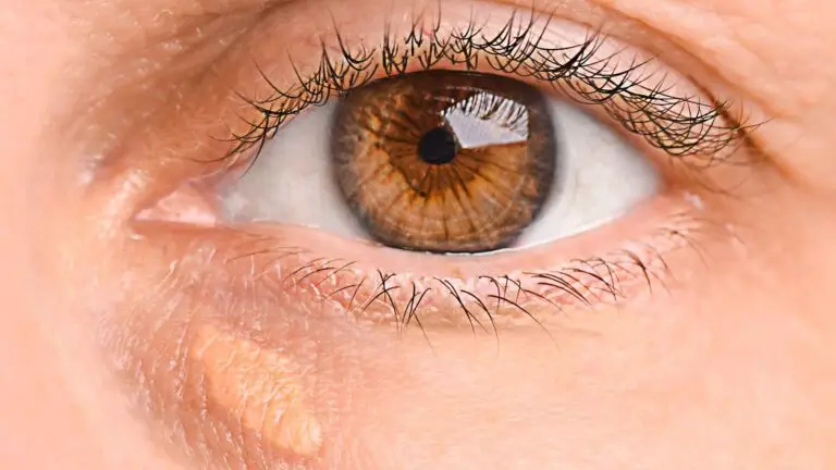
Retinal detachment is a serious eye condition that occurs when the retina – the light-sensitive layer at the back of the eye – separates from its supporting tissues. This separation deprives the retinal cells of oxygen and nourishment, which can lead to permanent vision loss if not promptly treated.
The Anatomy of the Eye
To understand retinal detachment, it’s helpful to know a bit about the eye’s structure:
- Retina: A thin layer of tissue lining the back of the eye, responsible for converting light into neural signals that the brain interprets as vision.
- Vitreous: A gel-like substance that fills the eye and helps maintain its shape.
- Choroid: The layer of blood vessels that supplies oxygen and nutrients to the retina.
When the retina detaches, it separates from the choroid, disrupting the normal visual process.

What Causes Retinal Detachment?
Retinal detachment can occur due to various reasons. Understanding these causes can help in prevention and early detection:
- Posterior Vitreous Detachment (PVD)
As we age, the vitreous gel in our eyes can shrink and pull away from the retina. While this is a normal part of ageing, sometimes it can lead to a tear in the retina, which can progress to a detachment.
- Trauma
Eye injuries or head trauma can cause the retina to detach. This is why protective eyewear is crucial in sports and certain work environments.
- High Myopia (Severe Short-sightedness)
People with high levels of myopia have elongated eyeballs, which can increase the risk of retinal detachment.
- Previous Eye Surgery
Although rare, complications from eye surgeries, including cataract surgery, can sometimes lead to retinal detachment.
- Genetic Factors
Some inherited eye conditions can increase the risk of retinal detachment.
- Diabetic Retinopathy
Advanced diabetic eye disease can lead to the growth of abnormal blood vessels, which can pull on the retina and cause detachment.
Signs and Symptoms of Retinal Detachment
Recognising the signs of retinal detachment is crucial for seeking timely treatment. If you experience any of the following symptoms, it’s imperative to contact Blue Fin Vision® or your nearest eye clinic immediately:
Early Warning Signs
- Sudden increase in floaters: These appear as small dark spots or squiggly lines in your vision.
- Flashes of light: Often described as lightning streaks in the side vision.
- A curtain or shadow moving across your field of vision: This can indicate that the retina is already detaching.
Progressive Symptoms
- Loss of peripheral vision: You may notice a dark shadow at the edges of your sight.
- Blurred vision: Your overall vision may become less clear.
- Straight lines appearing curved: This distortion can be a sign of macular involvement.
It’s important to note that retinal detachment is painless, which is why being aware of these visual symptoms is crucial.

Retinal Detachment Treatment
- Laser Surgery (Photocoagulation)
For small tears or holes in the retina:
- A laser is used to create small burns around the retinal tear.
- This forms scar tissue that seals the retina to the underlying tissue.
- It’s often used as a preventive measure when a tear is detected before detachment occurs.
- Cryopexy (Freezing Treatment)
Similar to laser surgery, but using extreme cold:
- A freezing probe is applied to the outer surface of the eye.
- This freezes the area around the retinal tear, creating scar tissue to seal the retina.

- Pneumatic Retinopexy
For certain types of detachments:
- A gas bubble is injected into the vitreous cavity.
- The bubble presses against the detached retina, pushing it back into place.
- The patient must maintain a specific head position for several days to keep the bubble in the right place.
- Laser or cryopexy is often used in conjunction to seal the retinal tear.

- Scleral Buckle Surgery
A more invasive procedure for larger detachments:
- A flexible band is placed around the eye to counteract the forces pulling the retina out of place.
- This band is not visible and remains permanently attached to the eye.
- Often combined with other techniques like cryopexy.

- Vitrectomy
For complex detachments:
- The vitreous gel is removed from the eye.
- The surgeon may use laser treatment to repair tears.
A gas or silicone oil bubble is inserted to hold the retina in place while it heals.
Recovery and Aftercare
Recovery from retinal detachment surgery varies depending on the procedure:
- Initial recovery: Vision is usually blurry for a few weeks to months.
- Follow-up appointments: Regular check-ups are crucial to monitor healing.
- Activity restrictions: Your surgeon will advise on limitations, which may include avoiding strenuous activities and air travel.
Things to Avoid with Retinal Detachment
If you’ve been diagnosed with or treated for retinal detachment, it’s important to:
- Avoid rubbing your eyes: This can put pressure on the healing retina.
- Limit rapid eye movements: Follow your doctor’s advice on eye exercises.
- Refrain from heavy lifting: This can increase eye pressure.
- Postpone air travel: Changes in air pressure can affect gas bubbles used in some treatments.
- Avoid swimming: Until your eye has fully healed.
- Limit screen time: Excessive use of digital devices can strain your eyes.
Prevention and Regular Check-ups
While not all cases of retinal detachment can be prevented, regular eye examinations can help detect early signs of retinal problems. At Blue Fin Vision®, we recommend:
- Annual eye exams: Especially for those at higher risk.
- Prompt attention to symptoms: Don’t ignore sudden changes in vision.
- Protective eyewear: Use appropriate eye protection during sports or hazardous activities.
- Managing underlying conditions: Such as diabetes, which can affect retinal health.

Why Choose Blue Fin Vision® for Retinal Care?
At Blue Fin Vision® Eye Clinic, we offer:
- Expert Care: Our team of experienced ophthalmologists specialises in retinal disorders.
- Advanced Technology: We use the latest diagnostic and treatment technologies.
- Personalised Treatment Plans: Each patient receives a tailored approach to their eye care.
- Comprehensive Support: From diagnosis through to aftercare, we’re with you every step of the way.
Conclusion
Retinal detachment is a serious condition that requires immediate medical attention. Understanding the causes, recognising the symptoms, and knowing the treatment options are crucial steps in protecting your vision. At Blue Fin Vision®, we’re committed to providing the highest standard of care for all retinal conditions.
If you’re experiencing any symptoms of retinal detachment or are due for an eye examination, don’t hesitate to contact our Harley Street clinic. Remember, when it comes to retinal detachment, early detection and treatment are key to preserving your vision.
Your sight is precious, and at Blue Fin Vision®, we’re dedicated to helping you maintain healthy eyes for life. Trust us for all your retinal care needs in London, Essex, or Hertfordshire.