- Medically Reviewed by: Mr Mfazo Hove, Consultant Ophthalmic Surgeon
- Author: Chris Dunnington
- Published: October 27, 2025
- Last Updated: May 1, 2026
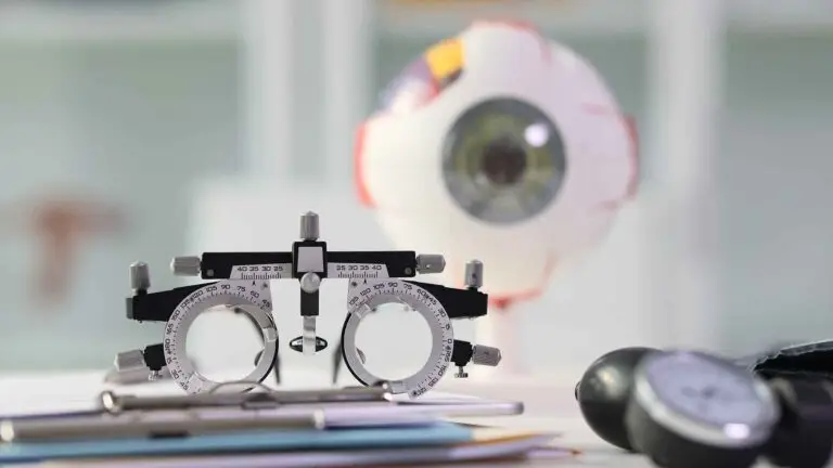
At Blue Fin Vision® Eye Clinic, the fundus photography test stands at the forefront of advanced eye diagnostics and testing – used daily by our top-rated private eye clinic team, including leading surgeons, top specialists, and renowned doctors. Understanding this essential test, how it works, and its purpose can empower patients to protect and optimise their vision with confidence.
The Gold Standard in Retinal Diagnostics
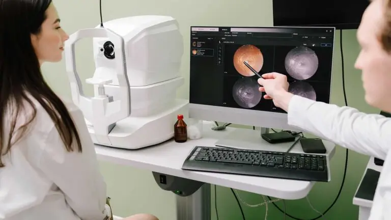
When it comes to maintaining eye health and detecting changes early, fundus photography is a cornerstone investigative tool. It enables clinicians to capture high-resolution colour images of the back of your eye, delivering a clear, detailed snapshot of the retina, optic nerve, and macula. Blue Fin Vision® utilises cutting-edge Zeiss technology, ensuring your test results set unsurpassed standards for clarity and precision.
We send all these test images and reports to patients with every consultation, allowing them to keep their own detailed record of eye health over time.

What Is a Fundus Photography Test?
A fundus photography test is a painless, non-invasive procedure in which an eye care specialist uses a sophisticated camera to take detailed photographs of your retina—the inner surface at the back of your eye. The fundus houses critical structures responsible for quality vision, such as the optic nerve, macula, and blood vessels. At Blue Fin Vision®, this procedure is performed at all clinic locations, leveraging world-class innovation for truly exceptional results.
What Does a Fundus Photography Eye Test Show?
Fundus photography is nothing short of revelatory when it comes to mapping and interpreting retinal health. The images it produces offer visual evidence of:
- Baseline appearance of the retina and optic nerve
- Blood vessels’ condition and branching patterns
- Subtle changes in the nerve fibre layer
- Early signs of retinal pathology, including haemorrhages, drusen (tiny yellow deposits), and pigmentary changes
By revealing these features, fundus photography allows for thorough screening and ongoing monitoring of progressive conditions, including age-related macular degeneration (AMD), diabetic retinopathy, and glaucoma.
Fundus Photography with Interpretation and Report

Fundus photo channels
This image shows four standard retinal fundus photographs taken using different light channels or filters to highlight various retinal structures. Here’s what each quadrant typically represents.
Top left:
- True‑colour fundus photograph RGB composite.
- Shows the retina as it appears in natural colour, highlighting the optic disc, macula, vessels, and any visible pathology for example, haemorrhages, drusen, scars.
- Used for general retinal assessment. For example, a supero-temporal choroidal naevus can be photographed here, allowing accurate yearly monitoring.
Top right:
- Red‑free image (green channel).
- Enhances contrast of blood vessels and nerve fibre layer by filtering out red light.
- Useful for detecting haemorrhages, microaneurysms, and RNFL defects.
Bottom left:
- Infrared (IR) image.
- Penetrates deeper retinal layers, visualising choroidal structures.
- Often used for macular and choroidal evaluation, especially before OCT scans.
Bottom right:
- Blue or autofluorescence (FAF) image.
- Detects naturally fluorescent compounds mainly lipofuscin in the RPE.
- Useful for identifying retinal pigment epithelial stress or damage for example in macular dystrophies and AMD.
In summary:
- Top left: Colour fundus photo
- Top right: Red‑free (green channel)
- Bottom left: Infrared
- Bottom right: Fundus autofluorescence (FAF)
Macular OCT report

This image shows a macular OCT (Optical Coherence Tomography) scan report specifically from an Optopol REVO FC device for the right eye (R). It provides a detailed, cross‑sectional and topographic analysis of the macula the central retina responsible for fine vision. For instance, the overlying OCT scan reveals there is no elevation of the retina above the choroidal naevus. The OCT report includes:
Top Left – Fundus Photo.
- A colour fundus image marking the scan area green box and scan line (green crosshair).
- It shows where the OCT cross‑section (B‑scan) has been taken.
Centre – OCT B‑scan (cross‑sectional image).
- A high‑resolution cross‑section through the retina at the macula.
- The red, blue, and cyan lines trace.
- Red: Inner Limiting Membrane (ILM), the top retinal surface.
- Cyan: Retinal Pigment Epithelium (RPE).
- Blue: IZ (Inner Segment /Outer Segment junction) or photoreceptor layer boundary.
- This allows measurement of total retinal and sub‑retinal thickness.
- The graph below shows retinal thickness along the scan line.
Bottom Left – ETDRS Thickness Map.
- Displays mean retinal thickness (in microns) in the standard ETDRS grid (Early Treatment Diabetic Retinopathy Study pattern).
- Central circle equals fovea.
- Inner and outer rings equal parafoveal and perifoveal zones.
- Example values:
- Central sector: 256 µm.
- Minimum in foveola: 219 µm.
- Mean macular thickness: 266 µm.
- Macular volume: 7.51 mm³.
- These are all within normal limits.
Top Right – Retina Significance Map.
- Compares retinal thickness to a normative database (age‑matched population).
- The colour scale (white‑pink‑red) indicates statistical deviation:
- Yellow or Red equals thinner or thicker than normal ranges.
- Green equals within normal range.
Middle Right – Retina Thickness Map.
- A colour topography of macular thickness.
- Blue equals thinner regions.
- Red equals thicker regions.
- The ETDRS grid is overlaid with numerical thickness values (µm).
Bottom Right – RPE Deformation Map.
- Assesses the contour of the retinal pigment epithelium (RPE) to detect distortions or elevations such as due to drusen, detachment, or sub‑RPE fluid.
- Colour‑coded scale:
- Blue equals normal or flat.
- Yellow to Red equals elevation or deformation.
Summary (bottom box).
- Minimum in Foveola: Thinnest point in macula, 219 µm.
- Central Sector: Average central 1 mm, 256 µm.
- Area Thickness: Mean across macula, 266 µm.
- Volume: Total macular volume, 7.51 mm³.

What Is the Purpose of Fundus Examination?
The fundamental purpose of fundus examination is to capture and record the health and structure of the retina, providing a vital baseline for all future retinal diagnostics and testing. These images help:
- Detect sight-threatening disease well before symptoms arise
- Guide treatment for retinal and optic nerve conditions
- Track progression and response to therapy with unparalleled accuracy
For Blue Fin Vision® patients, this translates into the reassurance of bespoke care plans, crafted with information gleaned from the latest technology by a team recognised at the very highest levels of UK healthcare.
Why Do I Need a Fundus Eye Test?
If you’ve been recommended a fundus eye test, you can be certain it’s for good reason. This scan is invaluable for:
- Early diagnosis of conditions that might otherwise go unnoticed until vision is threatened
- Tracking subtle changes that can only be seen with high-definition fundus imaging
- Providing top-rated, reliable evidence for specialists to customise your long-term treatment and prevention strategies
Many retinal problems, from age-related macular degeneration to diabetic changes, develop silently, and only sophisticated diagnostics reveal these changes in time for decisive intervention. Entrusting your fundus photography to Blue Fin Vision® guarantees meticulous standards carried out by the UK’s top doctors, all under one roof.
How Long Does the Fundus Photo Test Take?
Efficiency is another hallmark feature. A fundus photography test undertaken at Blue Fin Vision® typically lasts just a few minutes per eye, from positioning to image capture. Preparation is brief and results are available rapidly, enabling your consultant to discuss findings in real time and offer immediate advice. Most patients find the test swift and straightforward, without unnecessary waiting around.
Do You Need to Be Dilated for Fundus Photos?
Whether dilation is necessary depends on your individual case and which technology is deployed. Modern wide-field fundus cameras can often capture the required images without dilation, making the process even more convenient. In some instances, where the retina sits deep or subtle pathology is suspected, your specialist may recommend dilating the pupils to enhance photographic detail. The team will explain your options clearly and ensure you are comfortable throughout.
Is a Fundus Test Painful?
Rest easy: fundus photography is categorically non-invasive and well-tolerated. The procedure may involve a brief flash of light, which can feel mildly dazzling for a moment, but causes no discomfort. If dilation drops are used, they may lead to temporary light sensitivity or a fleeting blurring of vision, but the effects wear off rapidly. You’ll always be looked after by our compassionate team, whose focus is on keeping you comfortable and informed at every stage.
Blue Fin Vision®: Where Diagnostics Meet Distinction
Patients across London, Hertfordshire, and Essex choose Blue Fin Vision® for our advanced fundus photography, not just for the technology, but for our approach:
- Every scan is performed with award-winning equipment, ensuring nothing is missed.
- Our unified digital records mean instant access and seamless care across all clinics.
- Patient comfort and understanding remain our utmost priorities – every question answered, every procedure explained.
- Recognised at the Top Recommended level by Spears and Tatler, we are trusted leaders in high-spec vision diagnostics and testing.
Entrust Your Vision to Blue Fin Vision®
Choosing Blue Fin Vision® means choosing excellence, empathy, and innovation. If you’re considering a fundus photography test, or have been recommended one, arrange your appointment today at any of our state-of-the-art clinics in London, Hertfordshire, or Essex. Discover a higher standard of diagnostic eye care, personal attention, and consistency of service, always delivered by top-rated specialists whose every decision is driven by your vision’s best interests.
Your eyesight deserves nothing less than the expertise, technology, and assurance that Blue Fin Vision® delivers. Book your consultation now and experience the future of retinal diagnostics and testing.


