- Medically Reviewed by: Mr Mfazo Hove, Consultant Ophthalmic Surgeon
- Author: Chris Dunnington
- Published: November 10, 2025
- Last Updated: January 8, 2026
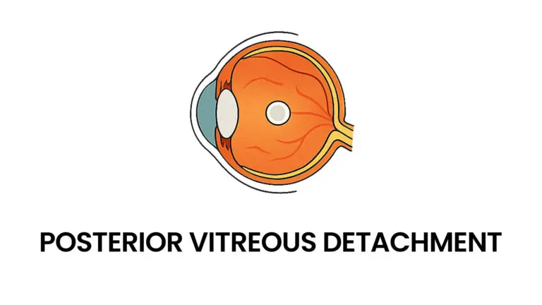
Posterior Vitreous Detachment, commonly known as PVD, is a natural age-related condition where the gel-like substance filling the eye, called the vitreous, begins to thin and pull away from the retina at the back of the eye. For many, PVD is a benign process that occurs gradually, often over several weeks or months, and does not lead to significant vision problems. However, it is a condition that warrants professional assessment, especially within a top-rated private eye clinic like Blue Fin Vision®, which is renowned for its leading surgeons and specialised care across London, Chelmsford, and Hatfield.
Causes of Posterior Vitreous Detachment
The primary cause of PVD is the ageing process. As part of normal ocular ageing, the vitreous gel gradually becomes more watery and less gel-like due to changes in collagen and water content. This process results in the vitreous shrinking and pulling away from the retina, the light-sensitive layer at the back of the eye.
Additional factors include:
- Refractive errors, particularly severe myopia (short-sightedness)
- Eye injuries or trauma
- Past eye surgeries, such as cataract removal
- Other conditions like lattice degeneration, which increase vitreoretinal adhesion, potentially complicating the detachment process
While PVD is typical over the age of 50, it can sometimes occur earlier, especially in individuals with high myopia or previous ocular trauma.
Symptoms of Posterior Vitreous Detachment
Understanding the symptoms is crucial for early detection and management. Most patients with PVD report:
- Floaters: Small, dark, shadowy shapes, strands, or cobweb-like shapes drifting across their vision
- Flashes of light: Often described as streaks or flickering lights, especially in dark environments
- A sudden increase in floaters or flashes, which can be alarming
In many cases, these symptoms resolve or become less noticeable over time. However, if the symptoms are sudden or persistent, it’s essential to seek prompt evaluation at a trusted private eye clinic. Rarely, PVD can lead to serious complications such as retinal tears or detachment, which could threaten vision if not treated swiftly.
How Serious is Posterior Vitreous Detachment?
While PVD itself is generally not dangerous and does not cause pain or permanent vision loss, its potential to lead to retinal tears or detachment makes it a condition that must be closely monitored by top specialists. Retinal detachment occurs in less than 15% of cases when PVD is coupled with vitreoretinal traction, but the risk remains significant enough to warrant urgent assessment if symptoms worsen.
Most patients experience gradual symptom resolution, with floaters and flashes diminishing over a few months. Nevertheless, understanding the signs of possible complications such as a sudden increase in floaters, flashing lights, or a shadow or curtain crossing the vision, and seeking immediate expert care is vital. For those in London, Chelmsford, or Hatfield, Blue Fin Vision® offers the reassurance of top doctors and leading surgeons who specialise in early diagnosis and safe, effective management.

Treatment Options for Posterior Vitreous Detachment
The good news is that in most cases, PVD requires no specific treatment because it usually resolves naturally. No eye exercises, dietary changes, or vitamins have proven effective in treating PVD.
However, in cases where complications such as retinal tears or detachment occur, options include:
- Laser treatment: To seal retinal tears and prevent detachment. This is a precise procedure performed by top surgeons at leading clinics.
- Vitrectomy: A more invasive surgical procedure where the vitreous gel is removed to repair retinal detachment. This is generally reserved for severe cases and carries risks such as infection or further retinal damage.
At Blue Fin Vision®, our team of top specialists utilises advanced diagnostic tools to detect any signs of retinal damage early. We tailor treatments to each patient’s specific needs, ensuring optimal safety and outcome.
Why Choose Blue Fin Vision®?
As a leading private eye clinic in the South East, Blue Fin Vision® is recognised for its excellence through peer awards, patient satisfaction, and collaborations with top-rated surgeons. Our centres in London, Chelmsford and Hatfield are staffed with the UK’s top ophthalmologists who specialise in retinal conditions, including PVD.
Patients benefit from:
- The latest diagnostic imaging, such as OCT and ultra-widefield retinal scans
- Personalised care guided by the UK’s top doctors and leading surgeons
- A seamless, surgeon-led network committed to consistency and excellence across all locations
- State-of-the-art technology, including laser and surgical options if required
- Clinical excellence, validated by Spears 500 and the Tatler Address Book
If you experience symptoms suggestive of PVD, such as floaters or flashes, don’t delay seeking expert advice. Early assessment is crucial, especially to rule out retinal tears or detachment that could threaten your eyesight.
Trust Your Vision to Blue Fin Vision®
Your eyes deserve nothing but the best. At Blue Fin Vision®, we understand the importance of maintaining clear, healthy vision and are committed to providing personalised, expert care in London, Chelmsford, and Hatfield. Our team of top specialists and surgeons deliver precise diagnosis, compassionate consultation, and exceptional treatment options.
Book your consultation today at the clinic most convenient to you – knowing your vision is in the hands of leading eye care professionals who prioritise your safety and satisfaction. Let us help you preserve your sight with patient-first care that truly sets the standard in modern ophthalmology.